3E9O | pdb_00003e9o

Crystal Structure of Yeast Prp8, Residues 1836-2092


Experimental Data Snapshot

  • Method: X-RAY DIFFRACTION
  • Resolution: 2.00 Å
  • R-Value Free: 
    0.245 (Depositor), 0.246 (DCC) 
  • R-Value Work: 
    0.177 (Depositor), 0.177 (DCC) 
  • R-Value Observed: 
    0.180 (Depositor) 

wwPDB Validation 3D Report Full Report

Validation slider image for 3E9O

This is version 1.2 of the entry. See complete history

Literature

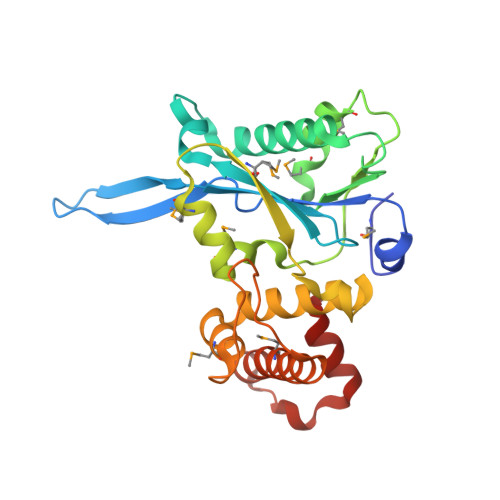
Structure and function of an RNase H domain at the heart of the spliceosome.

Pena, V.Rozov, A.Fabrizio, P.Luhrmann, R.Wahl, M.C.

(2008) EMBO J 27: 2929-2940

  • DOI: https://doi.org/10.1038/emboj.2008.209
  • Primary Citation Related Structures: 
    3E9L, 3E9O, 3E9P

  • PubMed Abstract: 

    Precursor-messenger RNA (pre-mRNA) splicing encompasses two sequential transesterification reactions in distinct active sites of the spliceosome that are transiently established by the interplay of small nuclear (sn) RNAs and spliceosomal proteins. Protein Prp8 is an active site component but the molecular mechanisms, by which it might facilitate splicing catalysis, are unknown. We have determined crystal structures of corresponding portions of yeast and human Prp8 that interact with functional regions of the pre-mRNA, revealing a phylogenetically conserved RNase H fold, augmented by Prp8-specific elements. Comparisons to RNase H-substrate complexes suggested how an RNA encompassing a 5'-splice site (SS) could bind relative to Prp8 residues, which on mutation, suppress splice defects in pre-mRNAs and snRNAs. A truncated RNase H-like active centre lies next to a known contact region of the 5'SS and directed mutagenesis confirmed that this centre is a functional hotspot. These data suggest that Prp8 employs an RNase H domain to help assemble and stabilize the spliceosomal catalytic core, coordinate the activities of other splicing factors and possibly participate in chemical catalysis of splicing.


  • Organizational Affiliation
    • Abteilung Zelluläre Biochemie, Max-Planck-Institut für Biophysikalische Chemie, Göttingen, Germany.

Macromolecule Content 

  • Total Structure Weight: 29.87 kDa 
  • Atom Count: 2,376 
  • Modeled Residue Count: 255 
  • Deposited Residue Count: 258 
  • Unique protein chains: 1

Macromolecules

Find similar proteins by:|  3D Structure
Entity ID: 1
MoleculeChains  Sequence LengthOrganismDetailsImage
Pre-mRNA-splicing factor 8258Saccharomyces cerevisiaeMutation(s): 0 
Gene Names: PRP8DBF3DNA39RNA8SLT21USA2YHR165C
UniProt
Find proteins for P33334 (Saccharomyces cerevisiae (strain ATCC 204508 / S288c))
Explore P33334 
Go to UniProtKB:  P33334
Entity Groups
Sequence Clusters30% Identity50% Identity70% Identity90% Identity95% Identity100% Identity
UniProt GroupP33334
Sequence Annotations
Expand
Reference Sequence

Small Molecules

Modified Residues  1 Unique
IDChains TypeFormula2D DiagramParent
MSE
Query on MSE
A
L-PEPTIDE LINKINGC5 H11 N O2 SeMET

Experimental Data & Validation

Experimental Data

  • Method: X-RAY DIFFRACTION
  • Resolution: 2.00 Å
  • R-Value Free:  0.245 (Depositor), 0.246 (DCC) 
  • R-Value Work:  0.177 (Depositor), 0.177 (DCC) 
  • R-Value Observed: 0.180 (Depositor) 
Space Group: P 31
Unit Cell:
Length ( Å )Angle ( ˚ )
a = 46.857α = 90
b = 46.857β = 90
c = 102.995γ = 120
Software Package:
Software NamePurpose
REFMACrefinement
DENZOdata reduction
SCALEPACKdata scaling
SHELXSphasing

Structure Validation

View Full Validation Report


Entry History 

Deposition Data

Revision History  (Full details and data files)

  • Version 1.0: 2008-10-07
    Type: Initial release
  • Version 1.1: 2011-07-13
    Changes: Advisory, Version format compliance
  • Version 1.2: 2024-10-30
    Changes: Data collection, Database references, Derived calculations, Structure summary