


Crystal structure of a putative oxidoreductase from Klebsiella pneumoniae
Eswaramoorthy, S., Mohammad, M.B., Thomas, C.A., Brown, A.C., Burley, S.K., Swaminathan, S.To be published.
Experimental Data Snapshot
wwPDB Validation 3D Report Full Report

Entity ID: 1 | |||||
|---|---|---|---|---|---|
| Molecule | Chains | Sequence Length | Organism | Details | Image |
| Putative oxidoreductase | A, B, C [auth D], D [auth E] | 364 | Klebsiella pneumoniae subsp. pneumoniae MGH 78578 | Mutation(s): 0 Gene Names: KPN78578_13420, KPN_01371 | 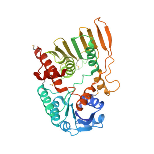 |
UniProt | |||||
Entity Groups | |||||
| Sequence Clusters | 30% Identity50% Identity70% Identity90% Identity95% Identity100% Identity | ||||
| UniProt Group | A6T882 | ||||
Sequence AnnotationsExpand | |||||
Reference Sequence | |||||
| Ligands 1 Unique | |||||
|---|---|---|---|---|---|
| ID | Chains | Name / Formula / InChI Key | 2D Diagram | 3D Interactions | |
| CL Download:Ideal Coordinates CCD File | E [auth A], F [auth B], G [auth D], H [auth E] | CHLORIDE ION Cl VEXZGXHMUGYJMC-UHFFFAOYSA-M |  | ||
| Modified Residues 1 Unique | |||||
|---|---|---|---|---|---|
| ID | Chains | Type | Formula | 2D Diagram | Parent |
| MSE Query on MSE | A, B, C [auth D], D [auth E] | L-PEPTIDE LINKING | C5 H11 N O2 Se |  | MET |
| Length ( Å ) | Angle ( ˚ ) |
|---|---|
| a = 52.91 | α = 90 |
| b = 79.75 | β = 94 |
| c = 209.144 | γ = 90 |
| Software Name | Purpose |
|---|---|
| CBASS | data collection |
| SHELX | model building |
| CNS | refinement |
| DENZO | data reduction |
| HKL-2000 | data scaling |
| SHELX | phasing |


