3E7K

Crystal Structure of an antiparallel coiled-coil tetramerization domain from TRPM7 channels


Experimental Data Snapshot

  • Method: X-RAY DIFFRACTION
  • Resolution: 2.01 Å
  • R-Value Free: 0.248 
  • R-Value Work: 0.188 
  • R-Value Observed: 0.191 

wwPDB Validation   3D Report Full Report


This is version 1.3 of the entry. See complete history


Literature

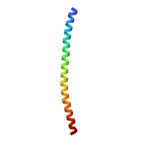
X-ray crystal structure of a TRPM assembly domain reveals an antiparallel four-stranded coiled-coil.

Fujiwara, Y.Minor, D.L.

(2008) J Mol Biol 383: 854-870

  • DOI: https://doi.org/10.1016/j.jmb.2008.08.059
  • Primary Citation of Related Structures:  
    3E7K

  • PubMed Abstract: 

    Transient receptor potential (TRP) channels comprise a large family of tetrameric cation-selective ion channels that respond to diverse forms of sensory input. Earlier studies showed that members of the TRPM subclass possess a self-assembling tetrameric C-terminal cytoplasmic coiled-coil domain that underlies channel assembly and trafficking. Here, we present the high-resolution crystal structure of the coiled-coil domain of the channel enzyme TRPM7. The crystal structure, together with biochemical experiments, reveals an unexpected four-stranded antiparallel coiled-coil architecture that bears unique features relative to other antiparallel coiled-coils. Structural analysis indicates that a limited set of interactions encode assembly specificity determinants and uncovers a previously unnoticed segregation of TRPM assembly domains into two families that correspond with the phylogenetic divisions seen for the complete subunits. Together, the data provide a framework for understanding the mechanism of TRPM channel assembly and highlight the diversity of forms found in the coiled-coil fold.


  • Organizational Affiliation

    Cardiovascular Research Institute, Departments of Biochemistry and Biophysics and Cellular and Molecular Pharmacology, California Institute for Quantitative Biosciences, University of California San Francisco, San Francisco, CA 94158-2330, USA.


Macromolecules
Find similar proteins by:  (by identity cutoff)  |  3D Structure
Entity ID: 1
MoleculeChains Sequence LengthOrganismDetailsImage
TRPM7 channel
A, B, C, D, E
A, B, C, D, E, F, G, H
56Rattus norvegicusMutation(s): 0 
UniProt
Find proteins for Q925B3 (Rattus norvegicus)
Explore Q925B3 
Go to UniProtKB:  Q925B3
Entity Groups  
Sequence Clusters30% Identity50% Identity70% Identity90% Identity95% Identity100% Identity
UniProt GroupQ925B3
Sequence Annotations
Expand
  • Reference Sequence
Experimental Data & Validation

Experimental Data

  • Method: X-RAY DIFFRACTION
  • Resolution: 2.01 Å
  • R-Value Free: 0.248 
  • R-Value Work: 0.188 
  • R-Value Observed: 0.191 
  • Space Group: C 1 2 1
Unit Cell:
Length ( Å )Angle ( ˚ )
a = 146.258α = 90
b = 35.674β = 124.46
c = 100.322γ = 90
Software Package:
Software NamePurpose
DENZOdata reduction
SCALEPACKdata scaling
PHASERphasing
REFMACrefinement
PDB_EXTRACTdata extraction
HKL-2000data reduction
HKL-2000data scaling

Structure Validation

View Full Validation Report


Entry History 

Deposition Data

Revision History  (Full details and data files)

  • Version 1.0: 2008-09-23
    Type: Initial release
  • Version 1.1: 2011-07-13
    Changes: Advisory, Version format compliance
  • Version 1.2: 2017-10-25
    Changes: Refinement description
  • Version 1.3: 2024-02-21
    Changes: Data collection, Database references