3E6Y | pdb_00003e6y

Structure of 14-3-3 in complex with the differentiation-inducing agent Cotylenin A


Experimental Data Snapshot

  • Method: X-RAY DIFFRACTION
  • Resolution: 2.50 Å
  • R-Value Free: 
    0.285 (Depositor), 0.295 (DCC) 
  • R-Value Work: 
    0.195 (Depositor), 0.209 (DCC) 
  • R-Value Observed: 
    0.200 (Depositor) 

Starting Model: experimental
View more details

wwPDB Validation 3D Report Full Report

Validation slider image for 3E6Y

Ligand Structure Quality Assessment 


This is version 1.4 of the entry. See complete history

Literature

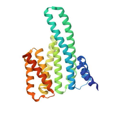
A structural rationale for selective stabilization of anti-tumor interactions of 14-3-3 proteins by cotylenin A

Ottmann, C.Weyand, M.Sassa, T.Inoue, T.Kato, N.Wittinghofer, A.Oecking, C.

(2009) J Mol Biology 386: 913-919

  • DOI: https://doi.org/10.1016/j.jmb.2009.01.005
  • Primary Citation Related Structures: 
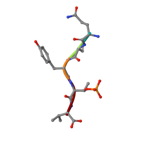
    3E6Y

  • PubMed Abstract: 

    Cotylenin A, a fungal metabolite originally described as a cytokinin-like bioactive substance against plants shows differentiation-inducing and anti-tumor activity in certain human cancers. Here, we present the crystal structure of cotylenin A acting on a 14-3-3 regulatory protein complex. By comparison with the closely related, but non-anticancer agent fusicoccin A, a rationale for the activity of cotylenin A in human cancers is presented. This class of fusicoccane diterpenoids are possible general modulators of 14-3-3 protein-protein interactions. In this regard, specificities for individual 14-3-3/target protein complexes might be achieved by varying the substituent pattern of the diterpene ring system. As the different activities of fusicoccin A and cotylenin A in human cancers suggest, hydroxylation of C12 might be a sufficient determinant of structural specificity.


  • Organizational Affiliation
    • Chemical Genomics Centre, Max Planck Society, Dortmund, Germany. christian.ottmann@cgc.mpg.de

Macromolecule Content 

  • Total Structure Weight: 61.49 kDa 
  • Atom Count: 3,926 
  • Modeled Residue Count: 476 
  • Deposited Residue Count: 530 
  • Unique protein chains: 2

Macromolecules

Find similar proteins by:|  3D Structure
Entity ID: 1
MoleculeChains  Sequence LengthOrganismDetailsImage
14-3-3-like protein C
A, B
260Nicotiana tabacumMutation(s): 0 
UniProt
Find proteins for P93343 (Nicotiana tabacum)
Explore P93343 
Go to UniProtKB:  P93343
Entity Groups
Sequence Clusters30% Identity50% Identity70% Identity90% Identity95% Identity100% Identity
UniProt GroupP93343
Sequence Annotations
Expand
Reference Sequence
Find similar proteins by:  Sequence   |   3D Structure  
Entity ID: 2
MoleculeChains  Sequence LengthOrganismDetailsImage
H+-ATPase phosphopeptide QSYpTV
C, D
5N/AMutation(s): 0 
UniProt
Find proteins for Q40409 (Nicotiana plumbaginifolia)
Explore Q40409 
Go to UniProtKB:  Q40409
Entity Groups
UniProt GroupQ40409
Sequence Annotations
Expand
Reference Sequence

Experimental Data & Validation

Experimental Data

  • Method: X-RAY DIFFRACTION
  • Resolution: 2.50 Å
  • R-Value Free:  0.285 (Depositor), 0.295 (DCC) 
  • R-Value Work:  0.195 (Depositor), 0.209 (DCC) 
  • R-Value Observed: 0.200 (Depositor) 
Space Group: P 1 21 1
Unit Cell:
Length ( Å )Angle ( ˚ )
a = 41.8α = 90
b = 141.9β = 114.1
c = 51.4γ = 90
Software Package:
Software NamePurpose
REFMACrefinement
XDSdata reduction
XSCALEdata scaling
MOLREPphasing

Structure Validation

View Full Validation Report


Ligand Structure Quality Assessment 


Entry History 

Deposition Data

Revision History  (Full details and data files)

  • Version 1.0: 2009-03-10
    Type: Initial release
  • Version 1.1: 2011-07-13
    Changes: Advisory, Version format compliance
  • Version 1.2: 2017-10-25
    Changes: Refinement description
  • Version 1.3: 2023-08-30
    Changes: Data collection, Database references, Derived calculations, Refinement description
  • Version 1.4: 2024-10-16
    Changes: Structure summary