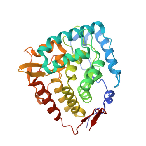
3E2T

The catalytic domain of chicken tryptophan hydroxylase 1 with bound tryptophan


Experimental Data Snapshot

  • Method: X-RAY DIFFRACTION
  • Resolution: 1.90 Å
  • R-Value Free: 0.226 
  • R-Value Work: 0.182 
  • R-Value Observed: 0.184 

Starting Model: experimental
View more details

wwPDB Validation   3D Report Full Report


Ligand Structure Quality Assessment 


This is version 1.2 of the entry. See complete history


Literature

Crystal structure of tryptophan hydroxylase with bound amino acid substrate

Windahl, M.S.Petersen, C.R.Christensen, H.E.M.Harris, P.

(2008) Biochemistry 47: 12087-12094

  • DOI: https://doi.org/10.1021/bi8015263
  • Primary Citation of Related Structures:  
    3E2T

  • PubMed Abstract: 

    Tryptophan hydroxylase (TPH) is a mononuclear non-heme iron enzyme, which catalyzes the reaction between tryptophan, O 2, and tetrahydrobiopterin (BH 4) to produce 5-hydroxytryptophan and 4a-hydroxytetrahydrobiopterin. This is the first and rate-limiting step in the biosynthesis of the neurotransmitter and hormone serotonin (5-hydroxytryptamine). We have determined the 1.9 A resolution crystal structure of the catalytic domain (Delta1-100/Delta415-445) of chicken TPH isoform 1 (TPH1) in complex with the tryptophan substrate and an iron-bound imidazole. This is the first structure of any aromatic amino acid hydroxylase with bound natural amino acid substrate. The iron coordination can be described as distorted trigonal bipyramidal coordination with His273, His278, and Glu318 (partially bidentate) and one imidazole as ligands. The tryptophan stacks against Pro269 with a distance of 3.9 A between the iron and the tryptophan Czeta3 atom that is hydroxylated. The binding of tryptophan and maybe the imidazole has caused the structural changes in the catalytic domain compared to the structure of the human TPH1 without tryptophan. The structure of chicken TPH1 is more compact, and the loops of residues Leu124-Asp139 and Ile367-Thr369 close around the active site. Similar structural changes are seen in the catalytic domain of phenylalanine hydroxylase (PAH) upon binding of substrate analogues norleucine and thienylalanine to the PAH.BH 4 complex. In fact, the chicken TPH1.Trp.imidazole structure resembles the PAH.BH 4.thienylalanine structure more (root-mean-square deviation for Calpha atoms of 0.90 A) than the human TPH1 structure (root-mean-square deviation of 1.47 A).


  • Organizational Affiliation

    Department of Basic Sciences and Environment, Faculty of Life Sciences, University of Copenhagen, Thorvaldsensvej 40, 1871 Frederiksberg C, Denmark.


Macromolecules
Find similar proteins by:  (by identity cutoff)  |  3D Structure
Entity ID: 1
MoleculeChains Sequence LengthOrganismDetailsImage
Tryptophan 5-hydroxylase 1314Gallus gallusMutation(s): 0 
EC: 1.14.16.4
UniProt
Find proteins for P70080 (Gallus gallus)
Explore P70080 
Go to UniProtKB:  P70080
Entity Groups  
Sequence Clusters30% Identity50% Identity70% Identity90% Identity95% Identity100% Identity
UniProt GroupP70080
Sequence Annotations
Expand
  • Reference Sequence
Small Molecules
Ligands 5 Unique
IDChains Name / Formula / InChI Key2D Diagram3D Interactions
TRP
Query on TRP

Download Ideal Coordinates CCD File 
E [auth A]TRYPTOPHAN
C11 H12 N2 O2
QIVBCDIJIAJPQS-VIFPVBQESA-N
PGE
Query on PGE

Download Ideal Coordinates CCD File 
F [auth A],
G [auth A]
TRIETHYLENE GLYCOL
C6 H14 O4
ZIBGPFATKBEMQZ-UHFFFAOYSA-N
SO4
Query on SO4

Download Ideal Coordinates CCD File 
C [auth A]SULFATE ION
O4 S
QAOWNCQODCNURD-UHFFFAOYSA-L
IMD
Query on IMD

Download Ideal Coordinates CCD File 
D [auth A]IMIDAZOLE
C3 H5 N2
RAXXELZNTBOGNW-UHFFFAOYSA-O
FE
Query on FE

Download Ideal Coordinates CCD File 
B [auth A]FE (III) ION
Fe
VTLYFUHAOXGGBS-UHFFFAOYSA-N
Experimental Data & Validation

Experimental Data

  • Method: X-RAY DIFFRACTION
  • Resolution: 1.90 Å
  • R-Value Free: 0.226 
  • R-Value Work: 0.182 
  • R-Value Observed: 0.184 
  • Space Group: C 2 2 21
Unit Cell:
Length ( Å )Angle ( ˚ )
a = 78α = 90
b = 155.3β = 90
c = 61.8γ = 90
Software Package:
Software NamePurpose
XSCALEdata scaling
MOLREPphasing
REFMACrefinement
PDB_EXTRACTdata extraction
ADSCdata collection
XDSdata reduction

Structure Validation

View Full Validation Report


Ligand Structure Quality Assessment 


Entry History 

Deposition Data

Revision History  (Full details and data files)

  • Version 1.0: 2008-11-04
    Type: Initial release
  • Version 1.1: 2011-07-13
    Changes: Version format compliance
  • Version 1.2: 2023-11-01
    Changes: Data collection, Database references, Derived calculations, Refinement description