3DXW | pdb_00003dxw

The crystal structure of alpha-amino-epsilon-caprolactam racemase from Achromobacter obae complexed with epsilon caprolactam


Experimental Data Snapshot

  • Method: X-RAY DIFFRACTION
  • Resolution: 2.41 Å
  • R-Value Free: 
    0.252 (Depositor), 0.249 (DCC) 
  • R-Value Work: 
    0.191 (Depositor), 0.191 (DCC) 
  • R-Value Observed: 
    0.194 (Depositor) 

Starting Model: experimental
View more details

wwPDB Validation 3D Report Full Report

Validation slider image for 3DXW

Ligand Structure Quality Assessment 


This is version 1.2 of the entry. See complete history

Literature

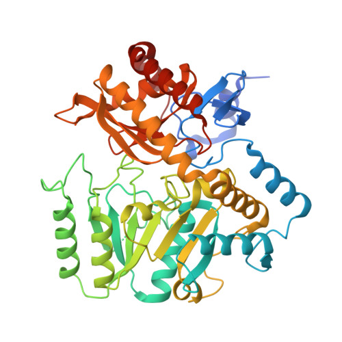
The novel structure of a pyridoxal 5'-phosphate-dependent fold-type I racemase, alpha-amino-epsilon-caprolactam racemase from Achromobacter obae

Okazaki, S.Suzuki, A.Mizushima, T.Kawano, T.Komeda, H.Asano, Y.Yamane, T.

(2009) Biochemistry 48: 941-950

  • DOI: https://doi.org/10.1021/bi801574p
  • Primary Citation Related Structures: 
    2ZUK, 3DXV, 3DXW

  • PubMed Abstract: 

    Alpha-amino-epsilon-caprolactam (ACL) racemase (ACLR) from Achromobacter obae catalyzes the interconversion of l- and d-ACL. ACLR belongs to the fold-type I group of pyridoxal 5'-phosphate (PLP) dependent enzymes. In this study, the first crystal structures of a fold-type I racemase are solved for the native form and epsilon-caprolactam-complexed form of ACLR at 2.21 and 2.40 A resolution, respectively. Based on the location of epsilon-caprolactam in the complex structure, the substrate-binding site is assigned between Trp49 and Tyr137. The carboxyl group of Asp210 is a reasonable candidate that recognizes the nitrogen atom of a lactam or amide in the substrate. Based on a structural comparison with fold-type III alanine racemase, Tyr137 is potentially the acid/base catalytic residue that is essential for the two-base racemization mechanism. The overall structure of ACLR is similar to that of fold-type I enzymes. A structural comparison with these enzymes explains the different reaction specificities.


  • Organizational Affiliation
    • Department of Biotechnology, Graduate School of Engineering, Nagoya University, Chikusa, Nagoya 464-8603, Japan.

Macromolecule Content 

  • Total Structure Weight: 95.84 kDa 
  • Atom Count: 6,374 
  • Modeled Residue Count: 848 
  • Deposited Residue Count: 904 
  • Unique protein chains: 1

Macromolecules

Find similar proteins by:|  3D Structure
Entity ID: 1
MoleculeChains  Sequence LengthOrganismDetailsImage
Alpha-amino-epsilon-caprolactam racemase
A, B
452Achromobacter obaeMutation(s): 0 
EC: 5.1 (PDB Primary Data), 5.1.1.15 (UniProt)
UniProt
Find proteins for Q7M181 (Achromobacter obae)
Explore Q7M181 
Go to UniProtKB:  Q7M181
Entity Groups
Sequence Clusters30% Identity50% Identity70% Identity90% Identity95% Identity100% Identity
UniProt GroupQ7M181
Sequence Annotations
Expand
Reference Sequence

Experimental Data & Validation

Experimental Data

  • Method: X-RAY DIFFRACTION
  • Resolution: 2.41 Å
  • R-Value Free:  0.252 (Depositor), 0.249 (DCC) 
  • R-Value Work:  0.191 (Depositor), 0.191 (DCC) 
  • R-Value Observed: 0.194 (Depositor) 
Space Group: P 1 21 1
Unit Cell:
Length ( Å )Angle ( ˚ )
a = 56.581α = 90
b = 60.653β = 103.07
c = 105.315γ = 90
Software Package:
Software NamePurpose
REFMACrefinement
HKL-2000data collection
HKL-2000data reduction
HKL-2000data scaling
MOLREPphasing

Structure Validation

View Full Validation Report


Ligand Structure Quality Assessment 


Entry History 

Deposition Data

Revision History  (Full details and data files)

  • Version 1.0: 2009-07-28
    Type: Initial release
  • Version 1.1: 2011-07-13
    Changes: Non-polymer description, Version format compliance
  • Version 1.2: 2023-11-01
    Changes: Data collection, Database references, Derived calculations, Refinement description