3DU0

E. coli dihydrodipicolinate synthase with first substrate, pyruvate, bound in active site


Experimental Data Snapshot

  • Method: X-RAY DIFFRACTION
  • Resolution: 2.00 Å
  • R-Value Free: 0.245 
  • R-Value Work: 0.188 
  • R-Value Observed: 0.191 

wwPDB Validation   3D Report Full Report


This is version 1.2 of the entry. See complete history


Literature

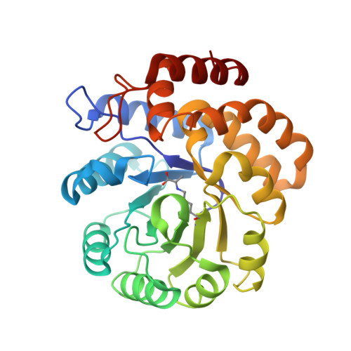
The high-resolution structure of dihydrodipicolinate synthase from Escherichia coli bound to its first substrate, pyruvate.

Devenish, S.R.Gerrard, J.A.Jameson, G.B.Dobson, R.C.

(2008) Acta Crystallogr Sect F Struct Biol Cryst Commun 64: 1092-1095

  • DOI: https://doi.org/10.1107/S1744309108033654
  • Primary Citation of Related Structures:  
    3DU0

  • PubMed Abstract: 

    Dihydrodipicolinate synthase (DHDPS) mediates the key first reaction common to the biosynthesis of (S)-lysine and meso-diaminopimelate, molecules which play a crucial cross-linking role in bacterial cell walls. An effective inhibitor of DHDPS would represent a useful antibacterial agent; despite extensive effort, a suitable inhibitor has yet to be found. In an attempt to examine the specificity of the active site of DHDPS, the enzyme was cocrystallized with the substrate analogue oxaloacetate. The resulting crystals diffracted to 2.0 A resolution, but solution of the protein structure revealed that pyruvate was bound in the active site rather than oxaloacetic acid. Kinetic analysis confirmed that the decarboxylation of oxaloacetate was not catalysed by DHDPS and was instead a slow spontaneous chemical process.


  • Organizational Affiliation

    School of Biological Sciences, University of Canterbury, Private Bag 4800, Christchurch 8020, New Zealand.


Macromolecules
Find similar proteins by:  (by identity cutoff)  |  3D Structure
Entity ID: 1
MoleculeChains Sequence LengthOrganismDetailsImage
Dihydrodipicolinate synthase
A, B
292Escherichia coli K-12Mutation(s): 0 
Gene Names: dapAb2478JW2463
EC: 4.2.1.52
UniProt
Find proteins for P0A6L2 (Escherichia coli (strain K12))
Explore P0A6L2 
Go to UniProtKB:  P0A6L2
Entity Groups  
Sequence Clusters30% Identity50% Identity70% Identity90% Identity95% Identity100% Identity
UniProt GroupP0A6L2
Sequence Annotations
Expand
  • Reference Sequence
Small Molecules
Ligands 3 Unique
IDChains Name / Formula / InChI Key2D Diagram3D Interactions
GOL
Query on GOL

Download Ideal Coordinates CCD File 
E [auth A],
F [auth A],
K [auth B]
GLYCEROL
C3 H8 O3
PEDCQBHIVMGVHV-UHFFFAOYSA-N
K
Query on K

Download Ideal Coordinates CCD File 
C [auth A],
D [auth A],
I [auth B],
J [auth B]
POTASSIUM ION
K
NPYPAHLBTDXSSS-UHFFFAOYSA-N
CL
Query on CL

Download Ideal Coordinates CCD File 
G [auth A],
H [auth A],
L [auth B],
M [auth B]
CHLORIDE ION
Cl
VEXZGXHMUGYJMC-UHFFFAOYSA-M
Modified Residues  1 Unique
IDChains TypeFormula2D DiagramParent
KPI
Query on KPI
A, B
L-PEPTIDE LINKINGC9 H16 N2 O4LYS
Experimental Data & Validation

Experimental Data

  • Method: X-RAY DIFFRACTION
  • Resolution: 2.00 Å
  • R-Value Free: 0.245 
  • R-Value Work: 0.188 
  • R-Value Observed: 0.191 
  • Space Group: P 31 2 1
Unit Cell:
Length ( Å )Angle ( ˚ )
a = 120.95α = 90
b = 120.95β = 90
c = 110.03γ = 120
Software Package:
Software NamePurpose
d*TREKdata scaling
MOLREPphasing
REFMACrefinement
PDB_EXTRACTdata extraction
d*TREKdata reduction

Structure Validation

View Full Validation Report


Entry History 

Deposition Data

Revision History  (Full details and data files)

  • Version 1.0: 2008-11-18
    Type: Initial release
  • Version 1.1: 2011-07-13
    Changes: Non-polymer description, Version format compliance
  • Version 1.2: 2019-12-18
    Changes: Advisory, Database references, Derived calculations