3DPC | pdb_00003dpc

Structure of E.coli Alkaline Phosphatase Mutant in Complex with a Phosphorylated Peptide


Experimental Data Snapshot

  • Method: X-RAY DIFFRACTION
  • Resolution: 2.30 Å
  • R-Value Free: 
    0.271 (Depositor), 0.250 (DCC) 
  • R-Value Work: 
    0.234 (Depositor), 0.239 (DCC) 

Starting Model: experimental
View more details

wwPDB Validation 3D Report Full Report

Validation slider image for 3DPC

This is version 1.5 of the entry. See complete history. 

Literature

Development of a universal phosphorylated peptide-binding protein for simultaneous assay of kinases

Li, W., Bi, L., Wang, W., Li, Y., Zhou, Y., Wei, H., Jiang, T., Bai, L., Chen, Y., Zhang, Z., Yuan, X., Xiao, J., Zhang, X.-E.

(2009) Biosens Bioelectron 24: 2871-2877

  • DOI: https://doi.org/10.1016/j.bios.2009.02.020
  • Primary Citation Related Structures: 
    3DPC

  • PubMed Abstract: 

    This study describes the development of a universal phosphorylated peptide-binding protein designed to simultaneously detect serine, threonine and tyrosine kinases. The Escherichia coli alkaline phosphatase (EAP) is a well-defined nonspecific phosphated monoesterase and Ser-, Thr- or Tyr-phosphorylated peptides served as substrates for EAP in preliminary experiments. Based on the known catalytic mechanism of EAP, the recombinant site-directed mutant EAP-S102L was generated, whose catalytic activity was blocked, but its binding ability was preserved. For EAP-S102L the catalytic rate constant, k(cat), was reduced by a factor of 1000, while the Michaelis-Menten constant, K(m), remained almost unchanged. Crystallographic analysis of the EAP-S102L/phophorylated peptide complex revealed that EAP-S102L could bind the phosphate group of the phosphorylated peptide but lacked nucleophilic attack potential which was essential for the catalytic ability of EAP. Finally, by combining the fluorescence-labeled EAP-S102L with non-phophorylated peptide chips, kinases could be detected from tumor cell samples. The recombinant EAP-S102L construct is perhaps the first functional binding protein derived from a native enzyme, illustrating how one single mutation tremendously alters protein function.


  • Organizational Affiliation: 
    • State Key Laboratory of Virology, Wuhan Institute of Virology, Chinese Academy of Sciences, Wuhan 430071, China.

Macromolecule Content 

  • Total Structure Weight: 97.29 kDa 
  • Atom Count: 6,921 
  • Modeled Residue Count: 907 
  • Deposited Residue Count: 920 
  • Unique protein chains: 2

Macromolecules

Find similar proteins by:|  3D Structure
Entity ID: 1
MoleculeChains  Sequence LengthOrganismDetailsImage
Alkaline phosphatase
A, B
455Escherichia coli K-12Mutation(s): 1 
Gene Names: phoA, b0383, JW0374
EC: 3.1.3.1
UniProt
Find proteins for P00634 (Escherichia coli (strain K12))
Explore P00634 
Go to UniProtKB:  P00634
Entity Groups
Sequence Clusters30% Identity50% Identity70% Identity90% Identity95% Identity100% Identity
UniProt GroupP00634
Sequence Annotations
Expand
Reference Sequence
Find similar proteins by:  Sequence   |   3D Structure  
Entity ID: 2
MoleculeChains  Sequence LengthOrganismDetailsImage
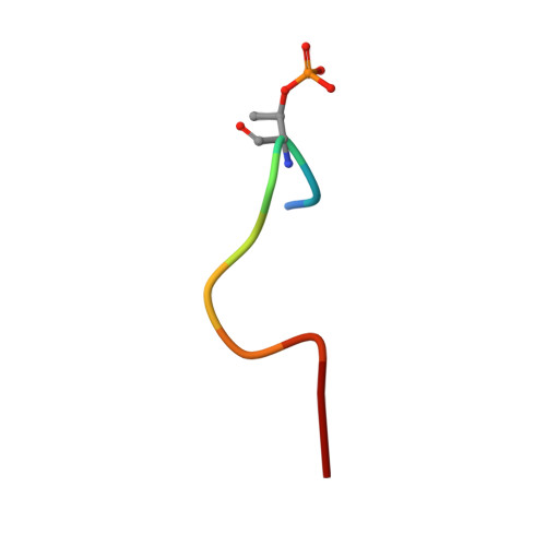
Phosphorylated Peptide10N/AMutation(s): 0 
Entity Groups
Sequence Clusters30% Identity50% Identity70% Identity90% Identity95% Identity100% Identity
Sequence Annotations
Expand
Reference Sequence

Small Molecules

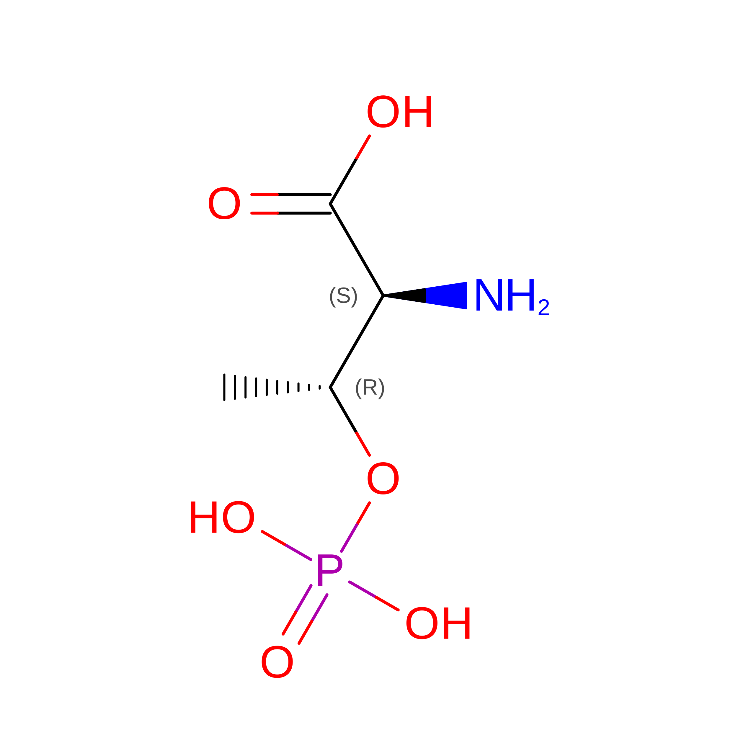
Modified Residues  1 Unique
IDChains TypeFormula2D DiagramParent
TPO
Query on TPO
C
L-PEPTIDE LINKINGC4 H10 N O6 PTHR

Experimental Data & Validation

Experimental Data

  • Method: X-RAY DIFFRACTION
  • Resolution: 2.30 Å
  • R-Value Free:  0.271 (Depositor), 0.250 (DCC) 
  • R-Value Work:  0.234 (Depositor), 0.239 (DCC) 
Space Group: P 21 21 21
Unit Cell:
Length ( Å )Angle ( ˚ )
a = 64.862α = 90
b = 107.74β = 90
c = 148.389γ = 90
Software Package:
Software NamePurpose
CrystalCleardata collection
PHASESphasing
CNSrefinement
d*TREKdata reduction
d*TREKdata scaling

Structure Validation

View Full Validation Report


Entry History 

Deposition Data

Revision History  (Full details and data files)

  • Version 1.0: 2009-06-16
    Type: Initial release
  • Version 1.1: 2011-07-13
    Changes: Source and taxonomy, Version format compliance
  • Version 1.2: 2014-02-05
    Changes: Derived calculations
  • Version 1.3: 2021-11-10
    Changes: Database references, Derived calculations
  • Version 1.4: 2023-11-01
    Changes: Data collection, Refinement description
  • Version 1.5: 2024-11-13
    Changes: Structure summary