3DOW

Complex structure of GABA type A receptor associated protein and its binding epitope on calreticulin


Experimental Data Snapshot

  • Method: X-RAY DIFFRACTION
  • Resolution: 2.30 Å
  • R-Value Free: 0.270 
  • R-Value Work: 0.232 
  • R-Value Observed: 0.233 

wwPDB Validation   3D Report Full Report


This is version 1.2 of the entry. See complete history


Literature

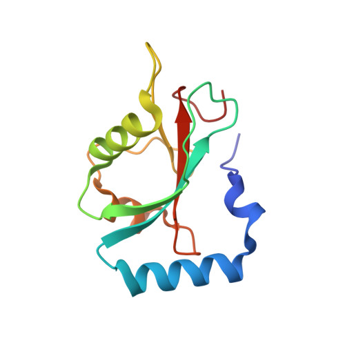
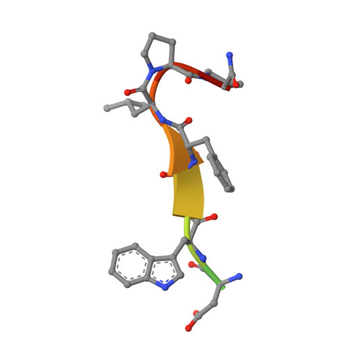
Structural framework of the GABARAP-calreticulin interface - implications for substrate binding to endoplasmic reticulum chaperones.

Thielmann, Y.Weiergraber, O.H.Mohrluder, J.Willbold, D.

(2009) FEBS J 276: 1140-1152

  • DOI: https://doi.org/10.1111/j.1742-4658.2008.06857.x
  • Primary Citation of Related Structures:  
    3DOW

  • PubMed Abstract: 

    The 4-aminobutyrate type A receptor-associated protein (GABARAP) is a versatile adaptor protein that plays an important role in intracellular vesicle trafficking, particularly in neuronal cells. We have investigated the structural determinants underlying the interaction of GABARAP with calreticulin using spectroscopic and crystallographic techniques. Specifically, we present the crystal structure of GABARAP in complex with its major binding epitope on the chaperone. Molecular modeling of a complex containing full-length calreticulin suggests a novel mode of substrate interaction, which may have functional implications for the calreticulin/calnexin family in general.


  • Organizational Affiliation

    Institut für Neurowissenschaften und Biophysik, Molekulare Biophysik, Forschungszentrum Jülich, Germany.


Macromolecules
Find similar proteins by:  (by identity cutoff)  |  3D Structure
Entity ID: 1
MoleculeChains Sequence LengthOrganismDetailsImage
Gamma-aminobutyric acid receptor-associated protein119Homo sapiensMutation(s): 0 
Gene Names: GABARAPFLC3B
UniProt & NIH Common Fund Data Resources
Find proteins for O95166 (Homo sapiens)
Explore O95166 
Go to UniProtKB:  O95166
PHAROS:  O95166
GTEx:  ENSG00000170296 
Entity Groups  
Sequence Clusters30% Identity50% Identity70% Identity90% Identity95% Identity100% Identity
UniProt GroupO95166
Sequence Annotations
Expand
  • Reference Sequence

Find similar proteins by:  Sequence   |   3D Structure  

Entity ID: 2
MoleculeChains Sequence LengthOrganismDetailsImage
CRT peptide12N/AMutation(s): 0 
UniProt & NIH Common Fund Data Resources
Find proteins for P27797 (Homo sapiens)
Explore P27797 
Go to UniProtKB:  P27797
PHAROS:  P27797
GTEx:  ENSG00000179218 
Entity Groups  
Sequence Clusters30% Identity50% Identity70% Identity90% Identity95% Identity100% Identity
UniProt GroupP27797
Sequence Annotations
Expand
  • Reference Sequence
Small Molecules
Ligands 1 Unique
IDChains Name / Formula / InChI Key2D Diagram3D Interactions
ZN
Query on ZN

Download Ideal Coordinates CCD File 
C [auth A]ZINC ION
Zn
PTFCDOFLOPIGGS-UHFFFAOYSA-N
Experimental Data & Validation

Experimental Data

  • Method: X-RAY DIFFRACTION
  • Resolution: 2.30 Å
  • R-Value Free: 0.270 
  • R-Value Work: 0.232 
  • R-Value Observed: 0.233 
  • Space Group: I 2 3
Unit Cell:
Length ( Å )Angle ( ˚ )
a = 97.043α = 90
b = 97.043β = 90
c = 97.043γ = 90
Software Package:
Software NamePurpose
MOSFLMdata reduction
SCALAdata scaling
PHENIXrefinement
PDB_EXTRACTdata extraction
ADSCdata collection
MOLREPphasing

Structure Validation

View Full Validation Report


Entry History 

Deposition Data

Revision History  (Full details and data files)

  • Version 1.0: 2009-02-24
    Type: Initial release
  • Version 1.1: 2011-07-13
    Changes: Version format compliance
  • Version 1.2: 2023-08-30
    Changes: Data collection, Database references, Derived calculations, Refinement description