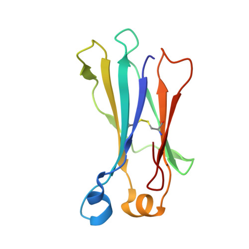
Structure of an isolated unglycosylated antibody C(H)2 domain.
Prabakaran, P., Vu, B.K., Gan, J., Feng, Y., Dimitrov, D.S., Ji, X.(2008) Acta Crystallogr D Biol Crystallogr 64: 1062-1067
- PubMed: 18931413 Search on PubMedSearch on PubMed Central
- DOI: https://doi.org/10.1107/S0907444908025274
- Primary Citation Related Structures:
3DJ9 - PubMed Abstract:
The C(H)2 (C(H)3 for IgM and IgE) domain of an antibody plays an important role in mediating effector functions and preserving antibody stability. It is the only domain in human immunoglobulins (Igs) which is involved in weak interchain protein-protein interactions with another C(H)2 domain solely through sugar moieties. The N-linked glycosylation at Asn297 is conserved in mammalian IgGs as well as in homologous regions of other antibody isotypes. To examine the structural details of the C(H)2 domain in the absence of glycosylation and other antibody domains, the crystal structure of an isolated unglycosylated antibody gamma1 C(H)2 domain was determined at 1.7 A resolution and compared with corresponding C(H)2 structures from intact Fc, IgG and Fc receptor complexes. Furthermore, the oligomeric state of the protein in solution was studied using size-exclusion chromatography. The results suggested that the unglycosylated human antibody C(H)2 domain is a monomer and that its structure is similar to that found in the intact Fc, IgG and Fc receptor complex structures. However, certain structural variations were observed in the Fc receptor-binding sites. Owing to its small size, stability and non-immunogenic Ig template, the C(H)2-domain structure could be useful for the development by protein design of antibody domains exerting effector functions and/or antigen specificity and as a robust scaffold in protein-engineering applications.
- Protein Interactions Group, Center for Cancer Research Nanobiology Program, National Cancer Institute, NIH, Frederick, MD 21702, USA.
Organizational Affiliation: