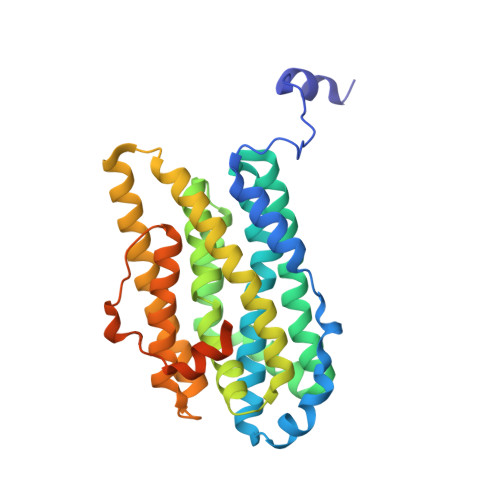
Structural and mutational studies of the carboxylate cluster in Iron-free Ribonucleotide Reductase R2.
Andersson, M.E., Hogbom, M., Rinaldo-Matthis, A., Blodig, W., Liang, Y., Persson, B.O., Sjoberg, B.M., Su, X.D., Nordlund, P.(2004) Biochemistry 43: 7966-7972
- PubMed: 15196041
- DOI: https://doi.org/10.1021/bi036088l
- Primary Citation of Related Structures:
3DHZ - PubMed Abstract:
The R2 protein of ribonucleotide reductase features a di-iron site deeply buried in the protein interior. The apo form of the R2 protein has an unusual clustering of carboxylate side chains at the empty metal-binding site. In a previous study, it was found that the loss of the four positive charge equivalents of the diferrous site in the apo protein appeared to be compensated for by the protonation of two histidine and two carboxylate side chains. We have studied the consequences of removing and introducing charged residues on the local hydrogen-bonding pattern in the region of the carboxylate cluster of Corynebacterium ammoniagenes and Escherichia coli protein R2 using site-directed mutagenesis and X-ray crystallography. The structures of the metal-free forms of wild-type C. ammoniagenes R2 and the mutant E. coli proteins D84N, S114D, E115A, H118A, and E238A have been determined and their hydrogen bonding and protonation states have been structurally assigned as far as possible. Significant alterations to the hydrogen-bonding patterns, protonation states, and hydration is observed for all mutant E. coli apo proteins as compared to wild-type apo R2. Further structural variations are revealed by the wild-type apo C. ammoniagenes R2 structure. The protonation and hydration effects seen in the carboxylate cluster appear to be due to two major factors: conservation of the overall charge of the site and the requirement of electrostatic shielding of clustered carboxylate residues. Very short hydrogen-bonding distances between some protonated carboxylate pairs are indicative of low-barrier hydrogen bonding.
- Department of Biochemistry and Biophysics, Stockholm University, SE-106 91 Stockholm, Sweden.
Organizational Affiliation:


