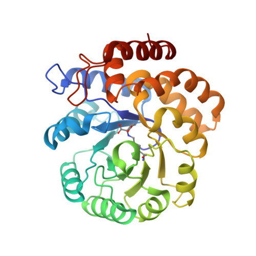
3DEN

Structure of E. coli DHDPS mutant Y107W


Experimental Data Snapshot

  • Method: X-RAY DIFFRACTION
  • Resolution: 2.20 Å
  • R-Value Free: 0.241 
  • R-Value Work: 0.183 
  • R-Value Observed: 0.186 

Starting Model: experimental
View more details

wwPDB Validation   3D Report Full Report


This is version 1.3 of the entry. See complete history


Literature

Mutating the tight-dimer interface of dihydrodipicolinate synthase disrupts the enzyme quaternary structure: toward a monomeric enzyme

Pearce, F.G.Dobson, R.C.J.Weber, A.Lane, L.A.McCammon, M.G.Squire, M.A.Perugini, M.A.Jameson, G.B.Robinson, C.V.Gerrard, J.A.

(2008) Biochemistry 47: 12108-12117

  • DOI: https://doi.org/10.1021/bi801094t
  • Primary Citation of Related Structures:  
    3DEN

  • PubMed Abstract: 

    Dihydrodipicolinate synthase (DHDPS) is a tetrameric enzyme that is the first enzyme unique to the ( S)-lysine biosynthetic pathway in plants and bacteria. Previous studies have looked at the important role of Tyr107, an amino acid residue located at the tight-dimer interface between two monomers, in participating in a catalytic triad of residues during catalysis. In this study, we examine the importance of this residue in determining the quaternary structure of the DHDPS enzyme. The Tyr107 residue was mutated to tryptophan, and structural, biophysical, and kinetic studies were carried out on the mutant enzyme. These revealed that while the solid-state structure of the mutant enzyme was largely unchanged, as judged by X-ray crystallography, it exists as a mixture of primarily monomer and tetramer in solution, as determined by analytical ultracentrifugation, size-exclusion chromatography, and mass spectrometry. The catalytic ability of the DHDPS enzyme was reduced by the mutation, which also allowed the adventitious binding of alpha-ketoglutarate to the active site. A reduction in the apparent melting temperature of the mutant enzyme was observed. Thus, the tetrameric quaternary structure of DHDPS is critical to controlling specificity, heat stability, and intrinsic activity.


  • Organizational Affiliation

    School of Biological Sciences, University of Canterbury, Private Bag 4800, Christchurch 8020, New Zealand.


Macromolecules
Find similar proteins by:  (by identity cutoff)  |  3D Structure
Entity ID: 1
MoleculeChains Sequence LengthOrganismDetailsImage
Dihydrodipicolinate synthase
A, B
292Escherichia coli K-12Mutation(s): 1 
Gene Names: dapA
EC: 4.2.1.52
UniProt
Find proteins for P0A6L2 (Escherichia coli (strain K12))
Explore P0A6L2 
Go to UniProtKB:  P0A6L2
Entity Groups  
Sequence Clusters30% Identity50% Identity70% Identity90% Identity95% Identity100% Identity
UniProt GroupP0A6L2
Sequence Annotations
Expand
  • Reference Sequence
Small Molecules
Ligands 3 Unique
IDChains Name / Formula / InChI Key2D Diagram3D Interactions
PO4
Query on PO4

Download Ideal Coordinates CCD File 
F [auth A],
I [auth B],
J [auth B]
PHOSPHATE ION
O4 P
NBIIXXVUZAFLBC-UHFFFAOYSA-K
GOL
Query on GOL

Download Ideal Coordinates CCD File 
D [auth A],
E [auth A],
G [auth A]
GLYCEROL
C3 H8 O3
PEDCQBHIVMGVHV-UHFFFAOYSA-N
K
Query on K

Download Ideal Coordinates CCD File 
C [auth A],
H [auth B]
POTASSIUM ION
K
NPYPAHLBTDXSSS-UHFFFAOYSA-N
Modified Residues  1 Unique
IDChains TypeFormula2D DiagramParent
KGC
Query on KGC
A, B
L-PEPTIDE LINKINGC11 H18 N2 O6LYS
Experimental Data & Validation

Experimental Data

  • Method: X-RAY DIFFRACTION
  • Resolution: 2.20 Å
  • R-Value Free: 0.241 
  • R-Value Work: 0.183 
  • R-Value Observed: 0.186 
  • Space Group: P 31 2 1
Unit Cell:
Length ( Å )Angle ( ˚ )
a = 121.196α = 90
b = 121.196β = 90
c = 110.565γ = 120
Software Package:
Software NamePurpose
REFMACrefinement
CrystalCleardata collection
d*TREKdata reduction
d*TREKdata scaling
AMoREphasing

Structure Validation

View Full Validation Report


Entry History 

Deposition Data

Revision History  (Full details and data files)

  • Version 1.0: 2008-11-25
    Type: Initial release
  • Version 1.1: 2011-07-13
    Changes: Non-polymer description, Version format compliance
  • Version 1.2: 2021-11-10
    Changes: Database references, Derived calculations
  • Version 1.3: 2023-11-01
    Changes: Data collection, Refinement description