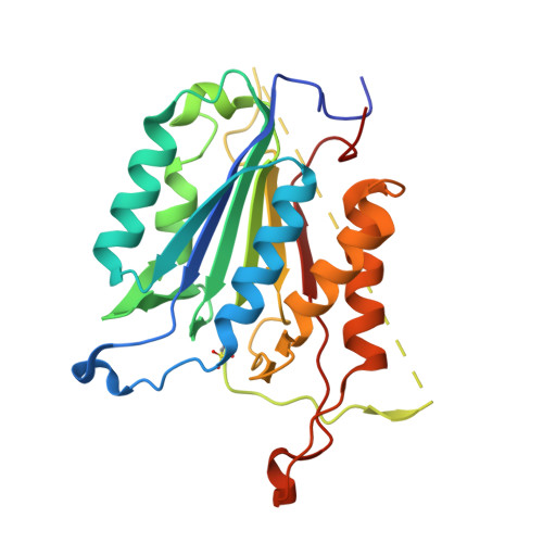
3DEJ

Crystal Structures of Caspase-3 with Bound Isoquinoline-1,3,4-trione Derivative Inhibitors


Experimental Data Snapshot

  • Method: X-RAY DIFFRACTION
  • Resolution: 2.60 Å
  • R-Value Free: 0.274 
  • R-Value Work: 0.214 
  • R-Value Observed: 0.217 

Starting Model: experimental
View more details

wwPDB Validation   3D Report Full Report


Ligand Structure Quality Assessment 


This is version 1.4 of the entry. See complete history


Literature

Isoquinoline-1,3,4-trione Derivatives Inactivate Caspase-3 by Generation of Reactive Oxygen Species

Du, J.-Q.Wu, J.Zhang, H.-J.Zhang, Y.-H.Qiu, B.-Y.Wu, F.Chen, Y.-H.Li, J.-Y.Nan, F.-J.Ding, J.-P.Li, J.

(2008) J Biol Chem 283: 30205-30215

  • DOI: https://doi.org/10.1074/jbc.M803347200
  • Primary Citation of Related Structures:  
    3DEH, 3DEI, 3DEJ, 3DEK

  • PubMed Abstract: 

    Caspase-3 is an attractive therapeutic target for treatment of diseases involving disregulated apoptosis. We report here the mechanism of caspase-3 inactivation by isoquinoline-1,3,4-trione derivatives. Kinetic analysis indicates the compounds can irreversibly inactivate caspase-3 in a 1,4-dithiothreitol (DTT)- and oxygen-dependent manner, implying that a redox cycle might take place in the inactivation process. Reactive oxygen species detection experiments using a chemical indicator, together with electron spin resonance measurement, suggest that ROS can be generated by reaction of isoquinoline-1,3,4-trione derivatives with DTT. Oxygen-free radical scavenger catalase and superoxide dismutase eliciting the inactivation of caspase-3 by the inhibitors confirm that ROS mediates the inactivation process. Crystal structures of caspase-3 in complexes with isoquinoline-1,3,4-trione derivatives show that the catalytic cysteine is oxidized to sulfonic acid (-SO(3)H) and isoquinoline-1,3,4-trione derivatives are bound at the dimer interface of caspase-3. Further mutagenesis study shows that the binding of the inhibitors with caspase-3 appears to be nonspecific. Isoquinoline-1,3,4-trione derivative-catalyzed caspase-3 inactivation could also be observed when DTT is substituted with dihydrolipoic acid, which exists widely in cells and might play an important role in the in vivo inactivation process in which the inhibitors inactivate caspase-3 in cells and then prevent the cells from apoptosis. These results provide valuable information for further development of small molecular inhibitors against caspase-3 or other oxidation-sensitive proteins.


  • Organizational Affiliation

    Chinese National Center for Drug Screening, Shanghai Institute of Materia Medica, Shanghai Institutes for Biological Sciences, Chinese Academy of Sciences, 189 Guo Shou Jing Road, Zhangjiang Hi-Tech Park, Shanghai 201203, People's Republic of China.


Macromolecules
Find similar proteins by:  (by identity cutoff)  |  3D Structure
Entity ID: 1
MoleculeChains Sequence LengthOrganismDetailsImage
Caspase-3
A, B, C, D
249Homo sapiensMutation(s): 0 
EC: 3.4.22.56
UniProt & NIH Common Fund Data Resources
Find proteins for P42574 (Homo sapiens)
Explore P42574 
Go to UniProtKB:  P42574
PHAROS:  P42574
GTEx:  ENSG00000164305 
Entity Groups  
Sequence Clusters30% Identity50% Identity70% Identity90% Identity95% Identity100% Identity
UniProt GroupP42574
Sequence Annotations
Expand
  • Reference Sequence
Small Molecules
Ligands 1 Unique
IDChains Name / Formula / InChI Key2D Diagram3D Interactions
RXC
Query on RXC

Download Ideal Coordinates CCD File 
E [auth C](1S)-1-(3-chlorophenyl)-2-oxo-2-[(1,3,4-trioxo-1,2,3,4-tetrahydroisoquinolin-5-yl)amino]ethyl acetate
C19 H13 Cl N2 O6
OVSAMUIBGQSLDC-INIZCTEOSA-N
Modified Residues  1 Unique
IDChains TypeFormula2D DiagramParent
OCS
Query on OCS
A, B, C, D
L-PEPTIDE LINKINGC3 H7 N O5 SCYS
Binding Affinity Annotations 
IDSourceBinding Affinity
RXC PDBBind:  3DEJ IC50: 10 (nM) from 1 assay(s)
Experimental Data & Validation

Experimental Data

  • Method: X-RAY DIFFRACTION
  • Resolution: 2.60 Å
  • R-Value Free: 0.274 
  • R-Value Work: 0.214 
  • R-Value Observed: 0.217 
  • Space Group: P 21 21 21
Unit Cell:
Length ( Å )Angle ( ˚ )
a = 65.433α = 90
b = 96.675β = 90
c = 180.446γ = 90
Software Package:
Software NamePurpose
DENZOdata reduction
SCALEPACKdata scaling
REFMACrefinement
PDB_EXTRACTdata extraction
HKL-2000data collection
HKL-2000data reduction
HKL-2000data scaling
CNSphasing

Structure Validation

View Full Validation Report


Ligand Structure Quality Assessment 


Entry History 

Deposition Data

Revision History  (Full details and data files)

  • Version 1.0: 2008-09-02
    Type: Initial release
  • Version 1.1: 2011-07-13
    Changes: Version format compliance
  • Version 1.2: 2017-10-25
    Changes: Refinement description
  • Version 1.3: 2023-11-01
    Changes: Data collection, Database references, Derived calculations, Refinement description
  • Version 1.4: 2023-11-15
    Changes: Data collection