3DDY

Structure of lumazine protein, an optical transponder of luminescent bacteria


Experimental Data Snapshot

  • Method: X-RAY DIFFRACTION
  • Resolution: 2.50 Å
  • R-Value Free: 0.250 
  • R-Value Work: 0.210 

wwPDB Validation   3D Report Full Report


This is version 2.0 of the entry. See complete history


Literature

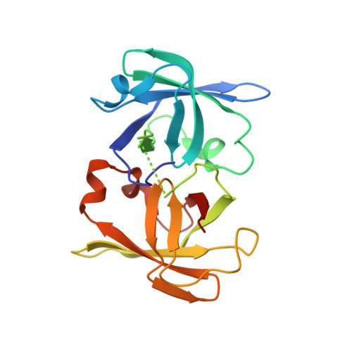
Structure of lumazine protein, an optical transponder of luminescent bacteria.

Chatwell, L.Illarionova, V.Illarionov, B.Eisenreich, W.Huber, R.Skerra, A.Bacher, A.Fischer, M.

(2008) J Mol Biol 382: 44-55

  • DOI: https://doi.org/10.1016/j.jmb.2008.06.052
  • Primary Citation of Related Structures:  
    3DDY

  • PubMed Abstract: 

    The intensely fluorescent lumazine protein is believed to be involved in the bioluminescence of certain marine bacteria. The sequence of the catalytically inactive protein resembles that of the enzyme riboflavin synthase. Its non-covalently bound fluorophore, 6,7-dimethyl-8-ribityllumazine, is the substrate of this enzyme and also the committed precursor of vitamin B(2). An extensive crystallization screen was performed using numerous single-site mutants of the lumazine protein from Photobacterium leiognathi in complex with its fluorophore and with riboflavin, respectively. Only the L49N mutant in complex with riboflavin yielded suitable crystals, allowing X-ray structure determination to a resolution of 2.5 A. The monomeric protein folds into two closely similar domains that are structurally related by pseudo-C(2) symmetry, whereby the entire domain topology resembles that of riboflavin synthase. Riboflavin is bound to a shallow cavity in the N-terminal domain of lumazine protein, whereas the C-terminal domain lacks a ligand.


  • Organizational Affiliation

    Lehrstuhl für Biologische Chemie, Technische Universität München, D-85350 Freising-Weihenstephan, Germany.


Macromolecules
Find similar proteins by:  (by identity cutoff)  |  3D Structure
Entity ID: 1
MoleculeChains Sequence LengthOrganismDetailsImage
Lumazine protein186Photobacterium leiognathi subsp. leiognathiMutation(s): 1 
Gene Names: lumP
UniProt
Find proteins for Q06877 (Photobacterium leiognathi)
Explore Q06877 
Go to UniProtKB:  Q06877
Entity Groups  
Sequence Clusters30% Identity50% Identity70% Identity90% Identity95% Identity100% Identity
UniProt GroupQ06877
Sequence Annotations
Expand
  • Reference Sequence
Small Molecules
Ligands 1 Unique
IDChains Name / Formula / InChI Key2D Diagram3D Interactions
RBF
Query on RBF

Download Ideal Coordinates CCD File 
B [auth A]RIBOFLAVIN
C17 H20 N4 O6
AUNGANRZJHBGPY-SCRDCRAPSA-N
Experimental Data & Validation

Experimental Data

  • Method: X-RAY DIFFRACTION
  • Resolution: 2.50 Å
  • R-Value Free: 0.250 
  • R-Value Work: 0.210 
  • Space Group: P 32 2 1
Unit Cell:
Length ( Å )Angle ( ˚ )
a = 79.188α = 90
b = 79.188β = 90
c = 67.62γ = 120
Software Package:
Software NamePurpose
MAR345dtbdata collection
PHASERphasing
CNSrefinement
MOSFLMdata reduction
SCALAdata scaling

Structure Validation

View Full Validation Report


Entry History 

Deposition Data

Revision History  (Full details and data files)

  • Version 1.0: 2008-07-01
    Type: Initial release
  • Version 1.1: 2011-07-13
    Changes: Non-polymer description, Version format compliance
  • Version 1.2: 2014-01-15
    Changes: Database references
  • Version 2.0: 2021-11-10
    Changes: Atomic model, Database references, Derived calculations