Crystal structures of Streptococcus suis mannonate dehydratase (ManD) and its complex with substrate: genetic and biochemical evidence for a catalytic mechanism
Zhang, Q., Gao, F., Peng, H., Cheng, H., Liu, Y., Tang, J., Thompson, J., Wei, G., Zhang, J., Du, Y., Yan, J., Gao, G.F.(2009) J Bacteriol 191: 5832-5837
- PubMed: 19617363 Search on PubMedSearch on PubMed Central
- DOI: https://doi.org/10.1128/JB.00599-09
- Primary Citation Related Structures:
3DBN, 3FVM - PubMed Abstract:
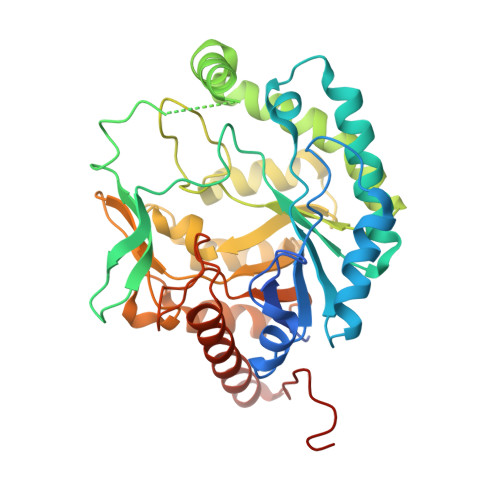
Mannonate dehydratase (ManD) is found only in certain bacterial species, where it participates in the dissimilation of glucuronate. ManD catalyzes the dehydration of d-mannonate to yield 2-keto-3-deoxygluconate (2-KDG), the carbon and energy source for growth. Selective inactivation of ManD by drug targeting is of therapeutic interest in the treatment of human Streptococcus suis infections. Here, we report the overexpression, purification, functional characterization, and crystallographic structure of ManD from S. suis. Importantly, by Fourier transform mass spectrometry, we show that 2-KDG is formed when the chemically synthesized substrate (d-mannonate) is incubated with ManD. Inductively coupled plasma-mass spectrometry revealed the presence of Mn(2+) in the purified protein, and in the solution state catalytically active ManD exists as a homodimer of two 41-kDa subunits. The crystal structures of S. suis ManD in native form and in complex with its substrate and Mn(2+) ion have been solved at a resolution of 2.9 A. The core structure of S. suis ManD is a TIM barrel similar to that of other members of the xylose isomerase-like superfamily. Structural analyses and comparative amino acid sequence alignments provide evidence for the importance of His311 and Tyr325 in ManD activity. The results of site-directed mutagenesis confirmed the functional role(s) of these residues in the dehydration reaction and a plausible mechanism for the ManD-catalyzed reaction is proposed.
- CAS Key Laboratory of Pathogenic Microbiology and Immunology, Institute of Microbiology, Chinese Academy of Sciences, Beijing 100101, China.
Organizational Affiliation: