3D9D

Nitroalkane oxidase: mutant D402N crystallized with 1-nitrohexane


Experimental Data Snapshot

  • Method: X-RAY DIFFRACTION
  • Resolution: 2.10 Å
  • R-Value Free: 0.219 
  • R-Value Work: 0.180 
  • R-Value Observed: 0.182 

wwPDB Validation   3D Report Full Report


Ligand Structure Quality Assessment 


This is version 1.4 of the entry. See complete history


Literature

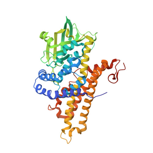
Crystal structures of intermediates in the nitroalkane oxidase reaction.

Heroux, A.Bozinovski, D.M.Valley, M.P.Fitzpatrick, P.F.Orville, A.M.

(2009) Biochemistry 48: 3407-3416

  • DOI: https://doi.org/10.1021/bi8023042
  • Primary Citation of Related Structures:  
    3D9D, 3D9E, 3D9F, 3D9G

  • PubMed Abstract: 

    The flavoenzyme nitroalkane oxidase is a member of the acyl-CoA dehydrogenase superfamily. Nitroalkane oxidase catalyzes the oxidation of neutral nitroalkanes to nitrite and the corresponding aldehydes or ketones. Crystal structures to 2.2 A resolution or better of enzyme complexes with bound substrates and of a trapped substrate-flavin adduct are described. The D402N enzyme has no detectable activity with neutral nitroalkanes [Valley, M. P., and Fitzpatrick, P. F. (2003) J. Am. Chem. Soc. 125, 8738-8739]. The structure of the D402N enzyme crystallized in the presence of 1-nitrohexane or 1-nitrooctane shows the presence of the substrate in the binding site. The aliphatic chain of the substrate extends into a tunnel leading to the enzyme surface. The oxygens of the substrate nitro group interact both with amino acid residues and with the 2'-hydroxyl of the FAD. When nitroalkane oxidase oxidizes nitroalkanes in the presence of cyanide, an electrophilic flavin imine intermediate can be trapped [Valley, M. P., Tichy, S. E., and Fitzpatrick, P. F. (2005) J. Am. Chem. Soc. 127, 2062-2066]. The structure of the enzyme trapped with cyanide during oxidation of 1-nitrohexane shows the presence of the modified flavin. A continuous hydrogen bond network connects the nitrogen of the CN-hexyl-FAD through the FAD 2'-hydroxyl to a chain of water molecules extending to the protein surface. Together, our complementary approaches provide strong evidence that the flavin cofactor is in the appropriate oxidation state and correlates well with the putative intermediate state observed within each of the crystal structures. Consequently, these results provide important structural descriptions of several steps along the nitroalkane oxidase reaction cycle.


  • Organizational Affiliation

    Department of Biology, Brookhaven National Laboratory, Upton, New York 11973, USA.


Macromolecules
Find similar proteins by:  (by identity cutoff)  |  3D Structure
Entity ID: 1
MoleculeChains Sequence LengthOrganismDetailsImage
Nitroalkane oxidase
A, B, C, D
438Fusarium oxysporumMutation(s): 1 
EC: 1.7.3.1
UniProt
Find proteins for Q8X1D8 (Fusarium oxysporum)
Explore Q8X1D8 
Go to UniProtKB:  Q8X1D8
Entity Groups  
Sequence Clusters30% Identity50% Identity70% Identity90% Identity95% Identity100% Identity
UniProt GroupQ8X1D8
Sequence Annotations
Expand
  • Reference Sequence
Small Molecules
Ligands 3 Unique
IDChains Name / Formula / InChI Key2D Diagram3D Interactions
FAD
Query on FAD

Download Ideal Coordinates CCD File 
E [auth A],
I [auth B],
M [auth C],
Q [auth D]
FLAVIN-ADENINE DINUCLEOTIDE
C27 H33 N9 O15 P2
VWWQXMAJTJZDQX-UYBVJOGSSA-N
N6C
Query on N6C

Download Ideal Coordinates CCD File 
F [auth A],
J [auth B],
N [auth C],
R [auth D]
1-nitrohexane
C6 H13 N O2
FEYJIFXFOHFGCC-UHFFFAOYSA-N
GOL
Query on GOL

Download Ideal Coordinates CCD File 
G [auth A]
H [auth A]
K [auth B]
L [auth B]
O [auth C]
G [auth A],
H [auth A],
K [auth B],
L [auth B],
O [auth C],
P [auth C],
S [auth D],
T [auth D]
GLYCEROL
C3 H8 O3
PEDCQBHIVMGVHV-UHFFFAOYSA-N
Experimental Data & Validation

Experimental Data

  • Method: X-RAY DIFFRACTION
  • Resolution: 2.10 Å
  • R-Value Free: 0.219 
  • R-Value Work: 0.180 
  • R-Value Observed: 0.182 
  • Space Group: P 32 2 1
Unit Cell:
Length ( Å )Angle ( ˚ )
a = 107.875α = 90
b = 107.875β = 90
c = 338.442γ = 120
Software Package:
Software NamePurpose
SCALAdata scaling
REFMACrefinement
PDB_EXTRACTdata extraction
CBASSdata collection
HKL-2000data reduction
HKL-2000data scaling
MOLREPphasing

Structure Validation

View Full Validation Report


Ligand Structure Quality Assessment 


Entry History 

Deposition Data

Revision History  (Full details and data files)

  • Version 1.0: 2009-04-07
    Type: Initial release
  • Version 1.1: 2011-07-13
    Changes: Non-polymer description, Version format compliance
  • Version 1.2: 2014-04-23
    Changes: Database references
  • Version 1.3: 2021-11-10
    Changes: Database references, Derived calculations
  • Version 1.4: 2023-11-01
    Changes: Data collection, Refinement description