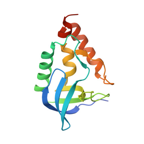
3D4W

Crystal structure of Staphylococcal nuclease variant Delta+PHS A109R at cryogenic temperature


Experimental Data Snapshot

  • Method: X-RAY DIFFRACTION
  • Resolution: 1.90 Å
  • R-Value Free: 0.252 
  • R-Value Work: 0.191 
  • R-Value Observed: 0.197 

Starting Model: experimental
View more details

wwPDB Validation   3D Report Full Report


This is version 1.5 of the entry. See complete history


Literature

Arginine residues at internal positions in a protein are always charged.

Harms, M.J.Schlessman, J.L.Sue, G.R.Garcia-Moreno E, B.

(2011) Proc Natl Acad Sci U S A 108: 18954-18959

  • DOI: https://doi.org/10.1073/pnas.1104808108
  • Primary Citation of Related Structures:  
    3D4W, 3D8G, 3DHQ

  • PubMed Abstract: 

    Many functionally essential ionizable groups are buried in the hydrophobic interior of proteins. A systematic study of Lys, Asp, and Glu residues at 25 internal positions in staphylococcal nuclease showed that their pK(a) values can be highly anomalous, some shifted by as many as 5.7 pH units relative to normal pK(a) values in water. Here we show that, in contrast, Arg residues at the same internal positions exhibit no detectable shifts in pK(a); they are all charged at pH ≤ 10. Twenty-three of these 25 variants with Arg are folded at both pH 7 and 10. The mean decrease in thermodynamic stability from substitution with Arg was 6.2 kcal/mol at this pH, comparable to that for substitution with Lys, Asp, or Glu at pH 7. The physical basis behind the remarkable ability of Arg residues to remain protonated in environments otherwise incompatible with charges is suggested by crystal structures of three variants showing how the guanidinium moiety of the Arg side chain is effectively neutralized through multiple hydrogen bonds to protein polar atoms and to site-bound water molecules. The length of the Arg side chain, and slight deformations of the protein, facilitate placement of the guanidinium moieties near polar groups or bulk water. This unique capacity of Arg side chains to retain their charge in dehydrated environments likely contributes toward the important functional roles of internal Arg residues in situations where a charge is needed in the interior of a protein, in a lipid bilayer, or in similarly hydrophobic environments.


  • Organizational Affiliation

    Department of Biophysics, Johns Hopkins University, 3400 North Charles Street, Baltimore, MD 21218, USA.


Macromolecules
Find similar proteins by:  (by identity cutoff)  |  3D Structure
Entity ID: 1
MoleculeChains Sequence LengthOrganismDetailsImage
Thermonuclease143Staphylococcus aureusMutation(s): 5 
Gene Names: nuc
EC: 3.1.31.1
UniProt
Find proteins for Q8NXI6 (Staphylococcus aureus (strain MW2))
Explore Q8NXI6 
Go to UniProtKB:  Q8NXI6
Entity Groups  
Sequence Clusters30% Identity50% Identity70% Identity90% Identity95% Identity100% Identity
UniProt GroupQ8NXI6
Sequence Annotations
Expand
  • Reference Sequence
Small Molecules
Ligands 2 Unique
IDChains Name / Formula / InChI Key2D Diagram3D Interactions
MPD
Query on MPD

Download Ideal Coordinates CCD File 
C [auth A](4S)-2-METHYL-2,4-PENTANEDIOL
C6 H14 O2
SVTBMSDMJJWYQN-YFKPBYRVSA-N
PO4
Query on PO4

Download Ideal Coordinates CCD File 
B [auth A]PHOSPHATE ION
O4 P
NBIIXXVUZAFLBC-UHFFFAOYSA-K
Experimental Data & Validation

Experimental Data

  • Method: X-RAY DIFFRACTION
  • Resolution: 1.90 Å
  • R-Value Free: 0.252 
  • R-Value Work: 0.191 
  • R-Value Observed: 0.197 
  • Space Group: P 21 21 21
Unit Cell:
Length ( Å )Angle ( ˚ )
a = 34.09α = 90
b = 59.843β = 90
c = 73.558γ = 90
Software Package:
Software NamePurpose
SAINTdata scaling
PHASERphasing
REFMACrefinement
PDB_EXTRACTdata extraction
APEXdata collection
SAINTdata reduction

Structure Validation

View Full Validation Report


Entry History 

Deposition Data

Revision History  (Full details and data files)

  • Version 1.0: 2009-01-06
    Type: Initial release
  • Version 1.1: 2011-07-13
    Changes: Version format compliance
  • Version 1.2: 2011-11-30
    Changes: Database references
  • Version 1.3: 2011-12-14
    Changes: Database references
  • Version 1.4: 2021-10-20
    Changes: Database references, Derived calculations
  • Version 1.5: 2023-08-30
    Changes: Data collection, Refinement description