Involvement of water in carbohydrate-protein binding: concanavalin A revisited.
Kadirvelraj, R., Foley, B.L., Dyekjaer, J.D., Woods, R.J.(2008) J Am Chem Soc 130: 16933-16942
- PubMed: 19053475
- DOI: https://doi.org/10.1021/ja8039663
- Primary Citation of Related Structures:
3D4K - PubMed Abstract:
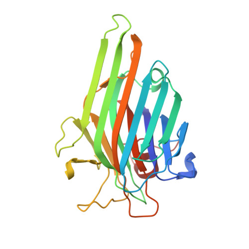
Ordered water molecules bound to protein surfaces, or in protein-ligand interfaces, are frequently observed by crystallography. The investigation of the impact of such conserved water molecules on protein stability and ligand affinity requires detailed structural, dynamic, and thermodynamic analyses. Several crystal structures of the legume lectin concanavalin A (Con A) bound to closely related carbohydrate ligands show the presence of a conserved water molecule that mediates ligand binding. Experimental thermodynamic and theoretical studies have examined the role of this conserved water in the complexation of Con A with a synthetic analog of the natural trisaccharide, in which a hydroxyethyl side chain replaces the hydroxyl group at the C-2 position in the central mannosyl residue. Molecular modeling earlier indicated (Clarke, C.; Woods, R. J.; Glushka, J.; Cooper, A.; Nutley, M. A.; Boons, G.-J. J. Am. Chem. Soc. 2001, 123, 12238-12247) that the hydroxyl group in this synthetic side chain could occupy a position equivalent to that of the conserved water, and thus might displace it. An interpretation of the experimental thermodynamic data, which was consistent with the displacement of the conserved water, was also presented. The current work reports the crystal structure of Con A with this synthetic ligand and shows that even though the position and interactions of the conserved water are distorted, this key water is not displaced by the hydroxyethyl moiety. This new structural data provides a firm basis for molecular dynamics simulations and thermodynamic integration calculations whose results indicate that differences in van der Waals contacts (insertion energy), rather than electrostatic interactions (charging energy) are fundamentally responsible for the lower affinity of the synthetic ligand. When combined with the new crystallographic data, this study provides a straightforward interpretation for the lower affinity of the synthetic analog; specifically, that it arises primarily from weaker interactions with the protein via the positionally perturbed conserved water. This interpretation is fully consistent with the experimental observations that the free energy of binding is enthalpy driven, that there is both less enthalpic gain and less entropic penalty for binding the synthetic ligand, relative to the natural trisaccharide, and that the entropic component does not arise from releasing an ordered water molecule from the protein surface to the bulk solvent.
- Complex Carbohydrate Research Center, University of Georgia, 315 Riverbend Road, Athens, Georgia 30602, USA.
Organizational Affiliation:


