3CZZ

Crystal structure of Cyanovirin-N domain B mutant


Experimental Data Snapshot

  • Method: X-RAY DIFFRACTION
  • Resolution: 1.36 Å
  • R-Value Free: 0.186 
  • R-Value Work: 0.165 
  • R-Value Observed: 0.165 

wwPDB Validation   3D Report Full Report


This is version 1.3 of the entry. See complete history


Literature

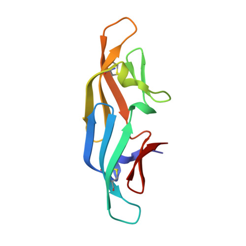
Solution and crystal structures of a sugar binding site mutant of cyanovirin-N: no evidence of domain swapping.

Matei, E.Furey, W.Gronenborn, A.M.

(2008) Structure 16: 1183-1194

  • DOI: https://doi.org/10.1016/j.str.2008.05.011
  • Primary Citation of Related Structures:  
    2RP3, 3CZZ

  • PubMed Abstract: 

    The cyanobacterial lectin Cyanovirin-N (CV-N) exhibits antiviral activity against HIV at a low nanomolar concentration by interacting with high-mannose oligosaccharides on the virus surface envelope glycoprotein gp120. Atomic structures of wild-type CV-N revealed a monomer in solution and a domain-swapped dimer in the crystal, with the monomer comprising two independent carbohydrate binding sites that individually bind with micromolar affinity to di- and trimannoses. In the mutant CVN(mutDB), the binding site on domain B was abolished and the protein was found to be completely inactive against HIV. We determined the solution NMR and crystal structures of this variant and characterized its sugar binding properties. In solution and the crystal, CVN(mutDB) is a monomer and no domain-swapping was observed. The protein binds to Man-3 and Man-9 with similar dissociation constants ( approximately 4 muM). This confirms that the nanomolar activity of wild-type CV-N is related to the multisite nature of the protein carbohydrate interaction.


  • Organizational Affiliation

    Department of Structural Biology, University of Pittsburgh School of Medicine, Pittsburgh, PA 15260, USA.


Macromolecules
Find similar proteins by:  (by identity cutoff)  |  3D Structure
Entity ID: 1
MoleculeChains Sequence LengthOrganismDetailsImage
Cyanovirin-N
A, B
101Nostoc ellipsosporumMutation(s): 6 
UniProt
Find proteins for P81180 (Nostoc ellipsosporum)
Explore P81180 
Go to UniProtKB:  P81180
Entity Groups  
Sequence Clusters30% Identity50% Identity70% Identity90% Identity95% Identity100% Identity
UniProt GroupP81180
Sequence Annotations
Expand
  • Reference Sequence
Experimental Data & Validation

Experimental Data

  • Method: X-RAY DIFFRACTION
  • Resolution: 1.36 Å
  • R-Value Free: 0.186 
  • R-Value Work: 0.165 
  • R-Value Observed: 0.165 
  • Space Group: P 21 21 21
Unit Cell:
Length ( Å )Angle ( ˚ )
a = 34.16α = 90
b = 58.02β = 90
c = 87.79γ = 90
Software Package:
Software NamePurpose
PHASERphasing
CNSrefinement
StructureStudiodata collection
d*TREKdata reduction
d*TREKdata scaling

Structure Validation

View Full Validation Report


Entry History 

Deposition Data

Revision History  (Full details and data files)

  • Version 1.0: 2008-08-19
    Type: Initial release
  • Version 1.1: 2011-07-13
    Changes: Version format compliance
  • Version 1.2: 2021-11-10
    Changes: Database references
  • Version 1.3: 2023-11-01
    Changes: Data collection, Refinement description