3CYT

REDOX CONFORMATION CHANGES IN REFINED TUNA CYTOCHROME C


Experimental Data Snapshot

  • Method: X-RAY DIFFRACTION
  • Resolution: 1.80 Å

wwPDB Validation   3D Report Full Report


This is version 2.0 of the entry. See complete history


Literature

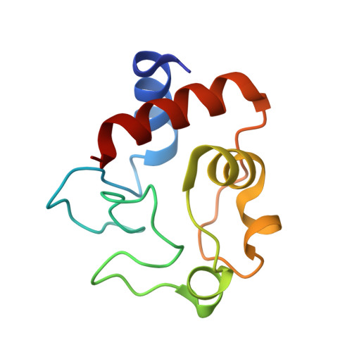
Redox conformation changes in refined tuna cytochrome c.

Takano, T.Dickerson, R.E.

(1980) Proc Natl Acad Sci U S A 77: 6371-6375

  • DOI: https://doi.org/10.1073/pnas.77.11.6371
  • Primary Citation of Related Structures:  
    3CYT

  • PubMed Abstract: 

    Tuna ferrocytochrome c and ferricytochrome c have been refined independently at high resolution (1.5 A and 1.8 A) to crystallographic residual errors of 17.3% and 20.8%, respectively. Small but significant conformational differences are seen surrounding a buried water molecule that is hydrogen bonded to Asn-52, Tyr-67, and Thr-78. In the oxidized state, this water molecule is 1.0 A closer to the heme and the heme has moved 0.15 A out of its heme crevice; both changes lead to a more polar microenvironment for the heme. Chemical modification studies, patterns of evolutionary conservatism, structural differences in bacterial cytochromes, and x-ray studies all agree that the "active site" for cytochrome c is bounded by lysines 8, 13,27, 72, 79, 86, and 87 (thus containing the evolutionary conservative 72-87 loop) and has the buried water molecule just below its surface and the opening of the heme crevice slightly to one side.


Macromolecules
Find similar proteins by:  (by identity cutoff)  |  3D Structure
Entity ID: 1
MoleculeChains Sequence LengthOrganismDetailsImage
CYTOCHROME CA [auth O],
B [auth I]
104Thunnus alalungaMutation(s): 0 
UniProt
Find proteins for P81459 (Thunnus alalunga)
Explore P81459 
Go to UniProtKB:  P81459
Entity Groups  
Sequence Clusters30% Identity50% Identity70% Identity90% Identity95% Identity100% Identity
UniProt GroupP81459
Sequence Annotations
Expand
  • Reference Sequence
Small Molecules
Ligands 1 Unique
IDChains Name / Formula / InChI Key2D Diagram3D Interactions
HEC
Query on HEC

Download Ideal Coordinates CCD File 
C [auth O],
D [auth I]
HEME C
C34 H34 Fe N4 O4
HXQIYSLZKNYNMH-LJNAALQVSA-N
Experimental Data & Validation

Experimental Data

  • Method: X-RAY DIFFRACTION
  • Resolution: 1.80 Å
  • Space Group: P 43
Unit Cell:
Length ( Å )Angle ( ˚ )
a = 74.42α = 90
b = 74.42β = 90
c = 36.3γ = 90

Structure Validation

View Full Validation Report


Entry History 

Deposition Data

  • Released Date: 1980-09-16 
  • Deposition Author(s): Takano, T.
  • This entry supersedes: 1CYT

Revision History  (Full details and data files)

  • Version 1.0: 1980-09-16
    Type: Initial release
  • Version 1.1: 2008-03-03
    Changes: Version format compliance
  • Version 1.2: 2011-07-13
    Changes: Version format compliance
  • Version 1.3: 2017-11-29
    Changes: Derived calculations, Other
  • Version 2.0: 2021-03-03
    Changes: Advisory, Atomic model, Data collection, Database references, Derived calculations, Non-polymer description, Structure summary