3CWS | pdb_00003cws

Crystal Structure of an AlkA Host/Guest Complex 2'-fluoro-2'-deoxyinosine:Thymine Base Pair


Experimental Data Snapshot

  • Method: X-RAY DIFFRACTION
  • Resolution: 2.30 Å
  • R-Value Free: 
    0.253 (Depositor) 
  • R-Value Work: 
    0.221 (Depositor), 0.218 (DCC) 

wwPDB Validation 3D Report Full Report

Validation slider image for 3CWS

This is version 1.3 of the entry. See complete history

Literature

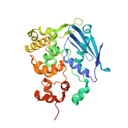
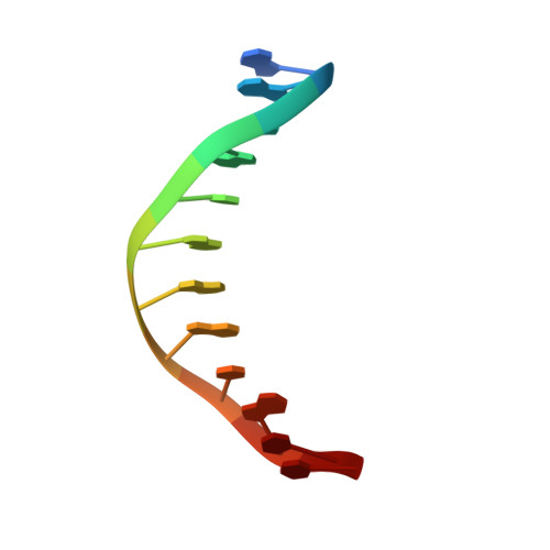
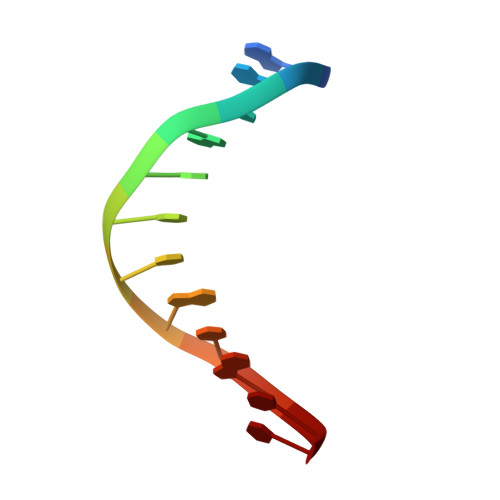
Structure of the E. coli DNA Glycosylase AlkA Bound to the Ends of Duplex DNA: A System for the Structure Determination of Lesion-Containing DNA.

Bowman, B.R.Lee, S.Wang, S.Verdine, G.L.

(2008) Structure 16: 1166-1174

  • DOI: https://doi.org/10.1016/j.str.2008.04.012
  • Primary Citation Related Structures: 
    3CVS, 3CVT, 3CW7, 3CWA, 3CWS, 3CWT, 3CWU

  • PubMed Abstract: 

    The constant attack on DNA by endogenous and exogenous agents gives rise to nucleobase modifications that cause mutations, which can lead to cancer. Visualizing the effects of these lesions on the structure of duplex DNA is key to understanding their biologic consequences. The most definitive method of obtaining such structures, X-ray crystallography, is troublesome to employ owing to the difficulty of obtaining diffraction-quality crystals of DNA. Here, we present a crystallization system that uses a protein, the DNA glycosylase AlkA, as a scaffold to mediate the crystallization of lesion-containing duplex DNA. We demonstrate the use of this system to facilitate the rapid structure determination of DNA containing the lesion 8-oxoguanine in several different sequence contexts, and also deoxyinosine and 1,N(6)-ethenoadenine, each stabilized as the corresponding 2'-flouro analog. The structures of 8-oxoguanine provide a correct atomic-level view of this important endogenous lesion in DNA.


  • Organizational Affiliation
    • Department of Chemistry and Chemical Biology, Harvard University, Cambridge, MA 02138, USA.

Macromolecule Content 

  • Total Structure Weight: 140.39 kDa 
  • Atom Count: 9,974 
  • Modeled Residue Count: 1,174 
  • Deposited Residue Count: 1,176 
  • Unique protein chains: 1
  • Unique nucleic acid chains: 2

Macromolecules


Find similar proteins by:|  3D Structure
Entity ID: 1
MoleculeChains  Sequence LengthOrganismDetailsImage
DNA-3-methyladenine glycosylase 2
A, B, C, D
282Escherichia coliMutation(s): 0 
Gene Names: alkAaidA
EC: 3.2.2.21
UniProt
Find proteins for P04395 (Escherichia coli (strain K12))
Explore P04395 
Go to UniProtKB:  P04395
Entity Groups
Sequence Clusters30% Identity50% Identity70% Identity90% Identity95% Identity100% Identity
UniProt GroupP04395
Sequence Annotations
Expand
Reference Sequence
Find similar nucleic acids by:  Sequence
Entity ID: 2
MoleculeChains LengthOrganismImage
DNA (5'-D(*DGP*DAP*DCP*DAP*DTP*DGP*DAP*(2FI)P*DTP*DGP*DCP*DC)-3')
E, G
12N/A
Sequence Annotations
Expand
Reference Sequence
Find similar nucleic acids by:  Sequence
Entity ID: 3
MoleculeChains LengthOrganismImage
DNA (5'-D(*DGP*DGP*DCP*DAP*DTP*DTP*DCP*DAP*DTP*DGP*DTP*DC)-3')
F, H
12N/A
Sequence Annotations
Expand
Reference Sequence

Experimental Data & Validation

Experimental Data

  • Method: X-RAY DIFFRACTION
  • Resolution: 2.30 Å
  • R-Value Free:  0.253 (Depositor) 
  • R-Value Work:  0.221 (Depositor), 0.218 (DCC) 
Space Group: P 1 21 1
Unit Cell:
Length ( Å )Angle ( ˚ )
a = 75.19α = 90
b = 100.988β = 94.03
c = 103.19γ = 90
Software Package:
Software NamePurpose
DENZOdata reduction
SCALEPACKdata scaling
CNSrefinement
PDB_EXTRACTdata extraction

Structure Validation

View Full Validation Report


Entry History 

Deposition Data

Revision History  (Full details and data files)

  • Version 1.0: 2008-09-02
    Type: Initial release
  • Version 1.1: 2011-07-13
    Changes: Version format compliance
  • Version 1.2: 2017-10-25
    Changes: Refinement description
  • Version 1.3: 2024-02-21
    Changes: Data collection, Database references, Derived calculations