3CV9 | pdb_00003cv9

Crystal structure of vitamin D hydroxylase cytochrome P450 105A1 (R73A/R84A mutant) in complex with 1alpha,25-dihydroxyvitamin D3


Experimental Data Snapshot

  • Method: X-RAY DIFFRACTION
  • Resolution: 1.70 Å
  • R-Value Free: 
    0.253 (Depositor), 0.254 (DCC) 
  • R-Value Work: 
    0.199 (Depositor), 0.200 (DCC) 
  • R-Value Observed: 
    0.202 (Depositor) 

Starting Model: experimental
View more details

wwPDB Validation 3D Report Full Report

Validation slider image for 3CV9

Ligand Structure Quality Assessment 


This is version 1.3 of the entry. See complete history

Literature

Structure-based design of a highly active vitamin D hydroxylase from Streptomyces griseolus CYP105A1

Hayashi, K.Sugimoto, H.Shinkyo, R.Yamada, M.Ikeda, S.Ikushiro, S.Kamakura, M.Shiro, Y.Sakaki, T.

(2008) Biochemistry 47: 11964-11972

  • DOI: https://doi.org/10.1021/bi801222d
  • Primary Citation Related Structures: 
    3CV8, 3CV9

  • PubMed Abstract: 

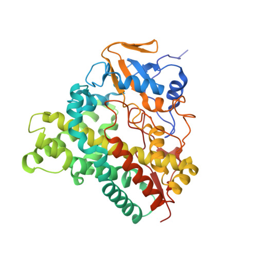
    CYP105A1 from Streptomyces griseolus has the capability of converting vitamin D 3 (VD 3) to its active form, 1alpha,25-dihydroxyvitamin D 3 (1alpha,25(OH) 2D 3) by a two-step hydroxylation reaction. Our previous structural study has suggested that Arg73 and Arg84 are key residues for the activities of CYP105A1. In this study, we prepared a series of single and double mutants by site-directed mutagenesis focusing on these two residues of CYP105A1 to obtain the hyperactive vitamin D 3 hydroxylase. R84F mutation altered the substrate specificity that gives preference to the 1alpha-hydroxylation of 25-hydroxyvitamin D 3 over the 25-hydroxylation of 1alpha-hydroxyvitamin D 3, opposite to the wild type and other mutants. The double mutant R73V/R84A exhibited 435- and 110-fold higher k cat/ K m values for the 25-hydroxylation of 1alpha-hydroxyvitamin D 3 and 1alpha-hydroxylation of 25-hydroxyvitamin D 3, respectively, compared with the wild-type enzyme. These values notably exceed those of CYP27A1, which is the physiologically essential VD 3 hydroxylase. Thus, we successfully generated useful enzymes of altered substrate preference and hyperactivity. Structural and kinetic analyses of single and double mutants suggest that the amino acid residues at positions 73 and 84 affect the location and conformation of the bound compound in the reaction site and those in the transient binding site, respectively.


  • Organizational Affiliation
    • Department of Biotechnology, Faculty of Engineering, Toyama Prefectural University, 5180 Kurokawa, Imizu, Toyama 939-0398, Japan.

Macromolecule Content 

  • Total Structure Weight: 45.94 kDa 
  • Atom Count: 3,686 
  • Modeled Residue Count: 401 
  • Deposited Residue Count: 412 
  • Unique protein chains: 1

Macromolecules

Find similar proteins by:|  3D Structure
Entity ID: 1
MoleculeChains  Sequence LengthOrganismDetailsImage
Cytochrome P450-SU1412Streptomyces griseolusMutation(s): 2 
Gene Names: CYP105A1
EC: 1.14.14.1 (PDB Primary Data), 1.14.15.22 (UniProt)
UniProt
Find proteins for P18326 (Streptomyces griseolus)
Explore P18326 
Go to UniProtKB:  P18326
Entity Groups
Sequence Clusters30% Identity50% Identity70% Identity90% Identity95% Identity100% Identity
UniProt GroupP18326
Sequence Annotations
Expand
Reference Sequence

Small Molecules

Ligands 2 Unique
IDChains Name / Formula / InChI Key2D Diagram3D Interactions
HEM

Query on HEM


Download:Ideal Coordinates CCD File
B [auth A]PROTOPORPHYRIN IX CONTAINING FE
C34 H32 Fe N4 O4
KABFMIBPWCXCRK-RGGAHWMASA-L
VDX

Query on VDX


Download:Ideal Coordinates CCD File
C [auth A]5-{2-[1-(5-HYDROXY-1,5-DIMETHYL-HEXYL)-7A-METHYL-OCTAHYDRO-INDEN-4-YLIDENE]-ETHYLIDENE}-4-METHYLENE-CYCLOHEXANE-1,3-DIOL
C27 H44 O3
GMRQFYUYWCNGIN-NKMMMXOESA-N

Experimental Data & Validation

Experimental Data

  • Method: X-RAY DIFFRACTION
  • Resolution: 1.70 Å
  • R-Value Free:  0.253 (Depositor), 0.254 (DCC) 
  • R-Value Work:  0.199 (Depositor), 0.200 (DCC) 
  • R-Value Observed: 0.202 (Depositor) 
Space Group: P 21 21 21
Unit Cell:
Length ( Å )Angle ( ˚ )
a = 53.315α = 90
b = 53.659β = 90
c = 140.605γ = 90
Software Package:
Software NamePurpose
REFMACrefinement
BSSdata collection
HKL-2000data reduction
HKL-2000data scaling
REFMACphasing

Structure Validation

View Full Validation Report


Ligand Structure Quality Assessment 


Entry History 

Deposition Data

Revision History  (Full details and data files)

  • Version 1.0: 2008-11-04
    Type: Initial release
  • Version 1.1: 2011-07-13
    Changes: Version format compliance
  • Version 1.2: 2021-11-10
    Changes: Database references, Derived calculations
  • Version 1.3: 2023-11-01
    Changes: Data collection, Refinement description