3CT4

Structure of Dha-kinase subunit DhaK from L. Lactis


Experimental Data Snapshot

  • Method: X-RAY DIFFRACTION
  • Resolution: 2.50 Å
  • R-Value Free: 0.267 
  • R-Value Work: 0.200 
  • R-Value Observed: 0.204 

wwPDB Validation   3D Report Full Report


This is version 1.4 of the entry. See complete history


Literature

X-ray Structures of the Three Lactococcus lactis Dihydroxyacetone Kinase Subunits and of a Transient Intersubunit Complex.

Zurbriggen, A.Jeckelmann, J.M.Christen, S.Bieniossek, C.Baumann, U.Erni, B.

(2008) J Biol Chem 283: 35789-35796

  • DOI: https://doi.org/10.1074/jbc.M804893200
  • Primary Citation of Related Structures:  
    3CR3, 3CT4, 3CT6

  • PubMed Abstract: 

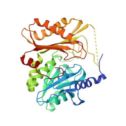
    Bacterial dihydroxyacetone (Dha) kinases do not exchange the ADP for ATP but utilize a subunit of the phosphoenolpyruvate carbohydrate phosphotransferase system for in situ rephosphorylation of a permanently bound ADP-cofactor. Here we report the 2.1-angstroms crystal structure of the transient complex between the phosphotransferase subunit DhaM of the phosphotransferase system and the nucleotide binding subunit DhaL of the Dha kinase of Lactococcus lactis, the 1.1-angstroms structure of the free DhaM dimer, and the 2.5-angstroms structure of the Dha-binding DhaK subunit. Conserved salt bridges and an edge-to-plane stacking contact between two tyrosines serve to orient DhaL relative to the DhaM dimer. The distance between the imidazole Nepsilon2 of the DhaM His-10 and the beta-phosphate oxygen of ADP, between which the gamma-phosphate is transferred, is 4.9 angstroms. An invariant arginine, which is essential for activity, is appropriately positioned to stabilize the gamma-phosphate in the transition state. The (betaalpha)4alpha fold of DhaM occurs a second time as a subfold in the DhaK subunit. By docking DhaL-ADP to this subfold, the nucleotide bound to DhaL and the C1-hydroxyl of Dha bound to DhaK are positioned for in-line transfer of phosphate.


  • Organizational Affiliation

    Departement für Chemie und Biochemie, Universität Bern, Freiestrasse 3, Bern CH-3012, Switzerland.


Macromolecules
Find similar proteins by:  (by identity cutoff)  |  3D Structure
Entity ID: 1
MoleculeChains Sequence LengthOrganismDetailsImage
PTS-dependent dihydroxyacetone kinase, dihydroxyacetone-binding subunit dhaK
A, B, C
332Lactococcus lactis subsp. lactisMutation(s): 0 
Gene Names: dhaK
EC: 2.7
UniProt
Find proteins for Q9CIV8 (Lactococcus lactis subsp. lactis (strain IL1403))
Explore Q9CIV8 
Go to UniProtKB:  Q9CIV8
Entity Groups  
Sequence Clusters30% Identity50% Identity70% Identity90% Identity95% Identity100% Identity
UniProt GroupQ9CIV8
Sequence Annotations
Expand
  • Reference Sequence
Experimental Data & Validation

Experimental Data

  • Method: X-RAY DIFFRACTION
  • Resolution: 2.50 Å
  • R-Value Free: 0.267 
  • R-Value Work: 0.200 
  • R-Value Observed: 0.204 
  • Space Group: P 31 2 1
Unit Cell:
Length ( Å )Angle ( ˚ )
a = 107.802α = 90
b = 107.802β = 90
c = 142.9γ = 120
Software Package:
Software NamePurpose
DENZOdata reduction
SCALEPACKdata scaling
PHENIXrefinement
PDB_EXTRACTdata extraction
SCALAdata scaling
PHASERphasing

Structure Validation

View Full Validation Report


Entry History 

Deposition Data

Revision History  (Full details and data files)

  • Version 1.0: 2008-10-21
    Type: Initial release
  • Version 1.1: 2011-07-13
    Changes: Version format compliance
  • Version 1.2: 2013-05-08
    Changes: Other
  • Version 1.3: 2017-10-25
    Changes: Refinement description
  • Version 1.4: 2023-08-30
    Changes: Data collection, Database references, Derived calculations, Refinement description, Structure summary