3CRE

Electron Microscopy model of the Saf Pilus- Type A


Experimental Data Snapshot

  • Method: ELECTRON MICROSCOPY
  • Resolution: 17.0 Å
  • Aggregation State: FILAMENT 
  • Reconstruction Method: HELICAL 

wwPDB Validation   3D Report Full Report


This is version 1.3 of the entry. See complete history


Literature

Structural analysis of the Saf pilus by electron microscopy and image processing.

Salih, O.Remaut, H.Waksman, G.Orlova, E.V.

(2008) J Mol Biol 379: 174-187

  • DOI: https://doi.org/10.1016/j.jmb.2008.03.056
  • Primary Citation of Related Structures:  
    3CRE, 3CRF

  • PubMed Abstract: 

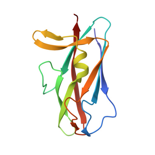
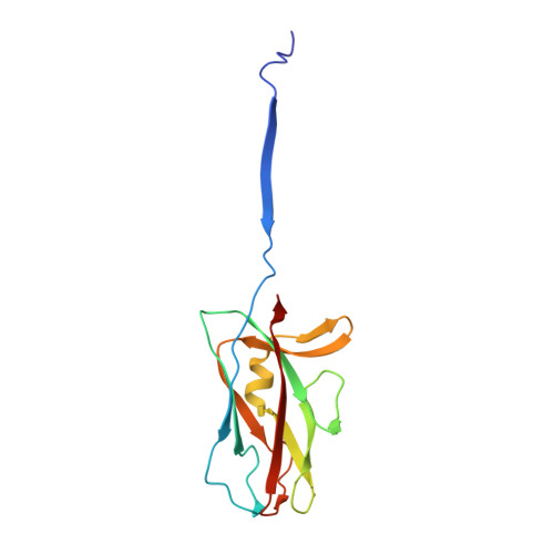
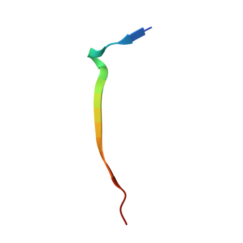
    Bacterial pili are important virulence factors involved in host cell attachment and/or biofilm formation, key steps in establishing and maintaining successful infection. Here we studied Salmonella atypical fimbriae (or Saf pili), formed by the conserved chaperone/usher pathway. In contrast to the well-established quaternary structure of typical/FGS-chaperone assembled, rod-shaped, chaperone/usher pili, little is known about the supramolecular organisation in atypical/FGL-chaperone assembled fimbriae. In our study, we have used negative stain electron microscopy and single-particle image analysis to determine the three-dimensional structure of the Salmonella typhimurium Saf pilus. Our results show atypical/FGL-chaperone assembled fimbriae are composed of highly flexible linear multi-subunit fibres that are formed by globular subunits connected to each other by short links giving a "beads on a string"-like appearance. Quantitative fitting of the atomic structure of the SafA pilus subunit into the electron density maps, in combination with linker modelling and energy minimisation, has enabled analysis of subunit arrangement and intersubunit interactions in the Saf pilus. Short intersubunit linker regions provide the molecular basis for flexibility of the Saf pilus by acting as molecular hinges allowing a large range of movement between consecutive subunits in the fibre.


  • Organizational Affiliation

    Institute of Structural Molecular Biology at UCL/Birkbeck, London WC1E 7HX, UK.


Macromolecules
Find similar proteins by:  (by identity cutoff)  |  3D Structure
Entity ID: 1
MoleculeChains Sequence LengthOrganismDetailsImage
Outer membrane protein123N/AMutation(s): 0 
Gene Names: safA
UniProt
Find proteins for Q8ZRK4 (Salmonella typhimurium (strain LT2 / SGSC1412 / ATCC 700720))
Explore Q8ZRK4 
Go to UniProtKB:  Q8ZRK4
Entity Groups  
Sequence Clusters30% Identity50% Identity70% Identity90% Identity95% Identity100% Identity
UniProt GroupQ8ZRK4
Sequence Annotations
Expand
  • Reference Sequence
Find similar proteins by:  (by identity cutoff)  |  3D Structure
Entity ID: 2
MoleculeChains Sequence LengthOrganismDetailsImage
Outer membrane protein144N/AMutation(s): 0 
Gene Names: safA
UniProt
Find proteins for Q8ZRK4 (Salmonella typhimurium (strain LT2 / SGSC1412 / ATCC 700720))
Explore Q8ZRK4 
Go to UniProtKB:  Q8ZRK4
Entity Groups  
Sequence Clusters30% Identity50% Identity70% Identity90% Identity95% Identity100% Identity
UniProt GroupQ8ZRK4
Sequence Annotations
Expand
  • Reference Sequence

Find similar proteins by:  Sequence   |   3D Structure  

Entity ID: 3
MoleculeChains Sequence LengthOrganismDetailsImage
Outer membrane protein19N/AMutation(s): 0 
UniProt
Find proteins for Q8ZRK4 (Salmonella typhimurium (strain LT2 / SGSC1412 / ATCC 700720))
Explore Q8ZRK4 
Go to UniProtKB:  Q8ZRK4
Entity Groups  
Sequence Clusters30% Identity50% Identity70% Identity90% Identity95% Identity100% Identity
UniProt GroupQ8ZRK4
Sequence Annotations
Expand
  • Reference Sequence
Experimental Data & Validation

Experimental Data

  • Method: ELECTRON MICROSCOPY
  • Resolution: 17.0 Å
  • Aggregation State: FILAMENT 
  • Reconstruction Method: HELICAL 
EM Software:
TaskSoftware PackageVersion
RECONSTRUCTIONIMAGIC

Structure Validation

View Full Validation Report


Entry History 

Deposition Data

Revision History  (Full details and data files)

  • Version 1.0: 2008-05-20
    Type: Initial release
  • Version 1.1: 2011-07-13
    Changes: Version format compliance
  • Version 1.2: 2018-07-18
    Changes: Data collection
  • Version 1.3: 2024-02-21
    Changes: Data collection, Database references, Refinement description