3CM7

Crystal Structure of XIAP-BIR3 domain in complex with Smac-mimetic compuond, Smac005


Experimental Data Snapshot

  • Method: X-RAY DIFFRACTION
  • Resolution: 3.10 Å
  • R-Value Free: 0.300 
  • R-Value Work: 0.233 
  • R-Value Observed: 0.236 

wwPDB Validation   3D Report Full Report


Ligand Structure Quality Assessment 


This is version 1.2 of the entry. See complete history


Literature

Targeting the X-linked inhibitor of apoptosis protein through 4-substituted azabicyclo[5.3.0]alkane smac mimetics. Structure, activity, and recognition principles.

Mastrangelo, E.Cossu, F.Milani, M.Sorrentino, G.Lecis, D.Delia, D.Manzoni, L.Drago, C.Seneci, P.Scolastico, C.Rizzo, V.Bolognesi, M.

(2008) J Mol Biol 384: 673-689

  • DOI: https://doi.org/10.1016/j.jmb.2008.09.064
  • Primary Citation of Related Structures:  
    3CLX, 3CM2, 3CM7

  • PubMed Abstract: 

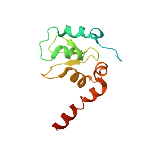
    The X-linked inhibitor of apoptosis protein (XIAP) is overexpressed in several malignant cells where it prevents apoptosis by binding to, and blocking, the activation of caspase-3, -7, and -9. Human XIAP (479 residues) is composed of three tandem-repeated baculoviral IAP repeat (BIR) domains (BIR1-3), and by a C-terminal RING domain. Smac-DIABLO [second mitochondria-derived activator of caspases (Smac)-direct IAP binding protein with low pI (DIABLO)], the natural antagonist of XIAP, binds through its N-terminal sequence AVPI to the same surface groove, in the BIR domains, that binds caspases. Synthetic compounds mimicking such tetrapeptide motif effectively block the interaction between IAP and active caspases, thus triggering apoptosis. Peptidomimetics based on an azabicyclo[x.y.0]alkane scaffolds, have been shown to bind the BIR3 domain of XIAP with micromolar to nanomolar affinities, thus presenting attractive features for drug lead optimization. Here we report a study on three newly synthesized Smac mimetics, which have been characterized in their complexes with XIAP BIR3 domain through X-ray crystallography and molecular modelling/docking simulations. Based on analysis of the crystal structures, we show that specific substitutions at the 4-position of the azabicyclo[5.3.0]alkane scaffold results in sizeable effects on the peptidomimetic-BIR3 domain affinity. By means of functional, biophysical and simulative approaches we also propose that the same Smac mimetics can bind XIAP BIR2 domain at a location structurally related to the BIR3 domain AVPI binding groove. Details of the XIAP-Smac mimetic recognition principles highlighted by this study are discussed in light of the drug-like profile of the three (potentially proapoptotic) compounds developed that show improved performance in ADMET (adsorption, distribution, metabolism, excretion and toxicity) tests.


  • Organizational Affiliation

    Department of Biomolecular Sciences and Biotechnology, CNR-INFM and CIMAINA, University of Milano, Via Celoria 26, I-20133 Milan, Italy.


Macromolecules
Find similar proteins by:  (by identity cutoff)  |  3D Structure
Entity ID: 1
MoleculeChains Sequence LengthOrganismDetailsImage
Baculoviral IAP repeat-containing protein 4A [auth C],
B [auth A],
C [auth B],
D
130Homo sapiensMutation(s): 0 
Gene Names: BIRC4API3IAP3XIAP
EC: 6.3.2
UniProt & NIH Common Fund Data Resources
Find proteins for P98170 (Homo sapiens)
Explore P98170 
Go to UniProtKB:  P98170
PHAROS:  P98170
GTEx:  ENSG00000101966 
Entity Groups  
Sequence Clusters30% Identity50% Identity70% Identity90% Identity95% Identity100% Identity
UniProt GroupP98170
Sequence Annotations
Expand
  • Reference Sequence
Small Molecules
Ligands 2 Unique
IDChains Name / Formula / InChI Key2D Diagram3D Interactions
X22
Query on X22

Download Ideal Coordinates CCD File 
F [auth C]
H [auth A]
J [auth B]
L [auth D]
M [auth D]
F [auth C],
H [auth A],
J [auth B],
L [auth D],
M [auth D],
N [auth D]
(3S,6S,7S,9aS)-6-{[(2S)-2-aminobutanoyl]amino}-N-(diphenylmethyl)-7-(hydroxymethyl)-5-oxooctahydro-1H-pyrrolo[1,2-a]azepine-3-carboxamide
C28 H36 N4 O4
WXYVFFGXHFXTLU-FCRIMTMASA-N
ZN
Query on ZN

Download Ideal Coordinates CCD File 
E [auth C],
G [auth A],
I [auth B],
K [auth D]
ZINC ION
Zn
PTFCDOFLOPIGGS-UHFFFAOYSA-N
Binding Affinity Annotations 
IDSourceBinding Affinity
X22 BindingDB:  3CM7 Ki: 250 (nM) from 1 assay(s)
IC50: min: 270, max: 290 (nM) from 2 assay(s)
PDBBind:  3CM7 IC50: 270 (nM) from 1 assay(s)
Experimental Data & Validation

Experimental Data

  • Method: X-RAY DIFFRACTION
  • Resolution: 3.10 Å
  • R-Value Free: 0.300 
  • R-Value Work: 0.233 
  • R-Value Observed: 0.236 
  • Space Group: P 41 21 2
Unit Cell:
Length ( Å )Angle ( ˚ )
a = 92.39α = 90
b = 92.39β = 90
c = 175.151γ = 90
Software Package:
Software NamePurpose
REFMACrefinement
MOSFLMdata reduction
SCALAdata scaling
MOLREPphasing

Structure Validation

View Full Validation Report


Ligand Structure Quality Assessment 


Entry History 

Deposition Data

Revision History  (Full details and data files)

  • Version 1.0: 2008-10-28
    Type: Initial release
  • Version 1.1: 2011-07-13
    Changes: Version format compliance
  • Version 1.2: 2023-08-30
    Changes: Data collection, Database references, Derived calculations, Refinement description