3CI3

Structure of the PduO-type ATP:co(I)rrinoid adenosyltransferase from Lactobacillus reuteri complexed with partial adenosylcobalamin and PPPi


Experimental Data Snapshot

  • Method: X-RAY DIFFRACTION
  • Resolution: 1.11 Å
  • R-Value Free: 0.180 
  • R-Value Work: 0.160 
  • R-Value Observed: 0.161 

wwPDB Validation   3D Report Full Report


Ligand Structure Quality Assessment 


This is version 1.3 of the entry. See complete history


Literature

Structural characterization of a human-type corrinoid adenosyltransferase confirms that coenzyme B12 is synthesized through a four-coordinate intermediate.

St Maurice, M.Mera, P.Park, K.Brunold, T.C.Escalante-Semerena, J.C.Rayment, I.

(2008) Biochemistry 47: 5755-5766

  • DOI: https://doi.org/10.1021/bi800132d
  • Primary Citation of Related Structures:  
    3CI1, 3CI3, 3CI4

  • PubMed Abstract: 

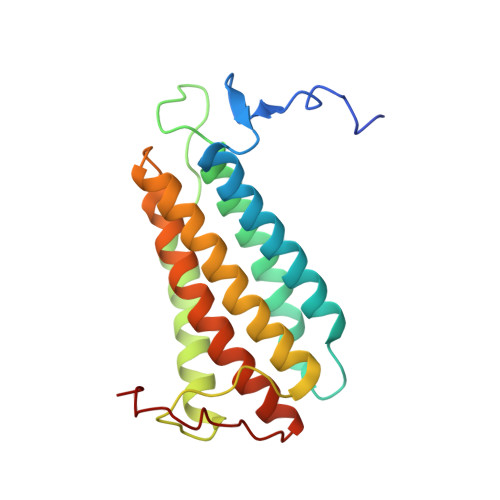
    ATP:cob(I)alamin adenosyltransferases (ACAs) catalyze the transfer of the 5'-deoxyadenosyl moiety from ATP to the upper axial ligand position of cobalamin in the synthesis of coenzyme B 12. For the ACA-catalyzed reaction to proceed, cob(II)alamin must be reduced to cob(I)alamin in the enzyme active site. This reduction is facilitated through the generation of a four-coordinate cob(II)alamin intermediate on the enzyme. We have determined the high-resolution crystal structure of a human-type ACA from Lactobacillus reuteri with a four-coordinate cob(II)alamin bound in the enzyme active site and with the product, adenosylcobalamin, partially occupied in the active site. The assembled structures represent snapshots of the steps in the ACA-catalyzed formation of the cobalt-carbon bond of coenzyme B 12. The structures define the corrinoid binding site and provide visual evidence for a base-off, four-coordinate cob(II)alamin intermediate. The complete structural description of ACA-mediated catalysis reveals the molecular features of four-coordinate cob(II)alamin stabilization and provides additional insights into the molecular basis for dysfunction in human patients suffering from methylmalonic aciduria.


  • Organizational Affiliation

    Departments of Biochemistry, University of Wisconsin, Madison, Wisconsin 53706, USA.


Macromolecules
Find similar proteins by:  (by identity cutoff)  |  3D Structure
Entity ID: 1
MoleculeChains Sequence LengthOrganismDetailsImage
Cobalamin adenosyltransferase PduO-like protein194Limosilactobacillus reuteriMutation(s): 0 
Gene Names: cobA
EC: 2.5.1.17
UniProt
Find proteins for Q50EJ2 (Limosilactobacillus reuteri)
Explore Q50EJ2 
Go to UniProtKB:  Q50EJ2
Entity Groups  
Sequence Clusters30% Identity50% Identity70% Identity90% Identity95% Identity100% Identity
UniProt GroupQ50EJ2
Sequence Annotations
Expand
  • Reference Sequence
Small Molecules
Ligands 5 Unique
IDChains Name / Formula / InChI Key2D Diagram3D Interactions
B12
Query on B12

Download Ideal Coordinates CCD File 
F [auth A]COBALAMIN
C62 H89 Co N13 O14 P
LKVIQTCSMMVGFU-DWSMJLPVSA-N
3PO
Query on 3PO

Download Ideal Coordinates CCD File 
D [auth A]TRIPHOSPHATE
H5 O10 P3
UNXRWKVEANCORM-UHFFFAOYSA-N
5AD
Query on 5AD

Download Ideal Coordinates CCD File 
E [auth A]5'-DEOXYADENOSINE
C10 H13 N5 O3
XGYIMTFOTBMPFP-KQYNXXCUSA-N
MG
Query on MG

Download Ideal Coordinates CCD File 
C [auth A]MAGNESIUM ION
Mg
JLVVSXFLKOJNIY-UHFFFAOYSA-N
NA
Query on NA

Download Ideal Coordinates CCD File 
B [auth A]SODIUM ION
Na
FKNQFGJONOIPTF-UHFFFAOYSA-N
Experimental Data & Validation

Experimental Data

  • Method: X-RAY DIFFRACTION
  • Resolution: 1.11 Å
  • R-Value Free: 0.180 
  • R-Value Work: 0.160 
  • R-Value Observed: 0.161 
  • Space Group: H 3
Unit Cell:
Length ( Å )Angle ( ˚ )
a = 67.965α = 90
b = 67.965β = 90
c = 110.941γ = 120
Software Package:
Software NamePurpose
REFMACrefinement
PDB_EXTRACTdata extraction
ADSCdata collection
HKL-2000data reduction
HKL-2000data scaling
MOLREPphasing

Structure Validation

View Full Validation Report


Ligand Structure Quality Assessment 


Entry History 

Deposition Data

Revision History  (Full details and data files)

  • Version 1.0: 2008-05-27
    Type: Initial release
  • Version 1.1: 2011-07-13
    Changes: Version format compliance
  • Version 1.2: 2012-10-24
    Changes: Non-polymer description
  • Version 1.3: 2024-02-21
    Changes: Data collection, Database references, Derived calculations