3CI0

The Crystal Structure of the GspK-GspI-GspJ complex from enterotoxigenic Escherichia coli Type 2 Secretion System


Experimental Data Snapshot

  • Method: X-RAY DIFFRACTION
  • Resolution: 2.20 Å
  • R-Value Free: 0.259 
  • R-Value Work: 0.192 
  • R-Value Observed: 0.195 

wwPDB Validation   3D Report Full Report


This is version 1.1 of the entry. See complete history


Literature

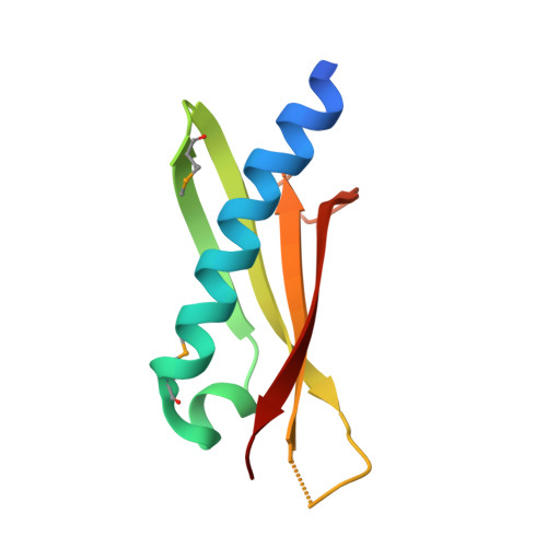
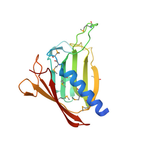
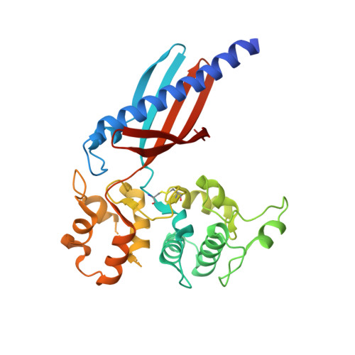
Structure of the GspK-GspI-GspJ complex from the enterotoxigenic Escherichia coli type 2 secretion system.

Korotkov, K.V.Hol, W.G.

(2008) Nat Struct Mol Biol 15: 462-468

  • DOI: https://doi.org/10.1038/nsmb.1426
  • Primary Citation of Related Structures:  
    3CI0

  • PubMed Abstract: 
  • Gram-negative bacteria translocate various proteins including virulence factors across their outer membrane via type 2 secretion systems (T2SSs). T2SSs are thought to contain a pseudopilus, a subcomplex formed by one major and several minor pseudopilins. We report the crystal structure of the complex formed by three minor pseudopilins from enterotoxigenic Escherichia coli ...

    Gram-negative bacteria translocate various proteins including virulence factors across their outer membrane via type 2 secretion systems (T2SSs). T2SSs are thought to contain a pseudopilus, a subcomplex formed by one major and several minor pseudopilins. We report the crystal structure of the complex formed by three minor pseudopilins from enterotoxigenic Escherichia coli. The GspK-GspI-GspJ complex has quasihelical characteristics and an architecture consistent with a localization at the pseudopilus tip. The alpha-domain of GspK has a previously unobserved fold with an unexpected dinuclear metal binding site. The area surrounding its disulfide bridge is conserved and might interact with other T2SS components or with secreted proteins.


    Organizational Affiliation

    Department of Biochemistry, Biomolecular Structure Center, University of Washington, Box 357742, Seattle, Washington 98195, USA.


Macromolecules
Find similar proteins by:  (by identity cutoff)  |  3D Structure
Entity ID: 1
MoleculeChainsSequence LengthOrganismDetailsImage
Pseudopilin GspIA [auth I]89Escherichia coliMutation(s): 0 
Gene Names: gspI
UniProt
Find proteins for Q8VPC3 (Escherichia coli)
Explore Q8VPC3 
Go to UniProtKB:  Q8VPC3
Entity Groups  
Sequence Clusters30% Identity50% Identity70% Identity90% Identity95% Identity100% Identity
UniProt GroupQ8VPC3
Protein Feature View
Expand
  • Reference Sequence
Find similar proteins by:  (by identity cutoff)  |  3D Structure
Entity ID: 2
MoleculeChainsSequence LengthOrganismDetailsImage
Pseudopilin GspJB [auth J]163Escherichia coliMutation(s): 0 
Gene Names: gspJBvCms2454_02423
UniProt
Find proteins for A0A4C3GMC1 (Escherichia coli)
Explore A0A4C3GMC1 
Go to UniProtKB:  A0A4C3GMC1
Entity Groups  
Sequence Clusters30% Identity50% Identity70% Identity90% Identity95% Identity100% Identity
UniProt GroupA0A4C3GMC1
Protein Feature View
Expand
  • Reference Sequence
Find similar proteins by:  (by identity cutoff)  |  3D Structure
Entity ID: 3
MoleculeChainsSequence LengthOrganismDetailsImage
Pseudopilin GspKC [auth K]298Escherichia coliMutation(s): 0 
Gene Names: gspK
UniProt
Find proteins for Q8VLK7 (Escherichia coli)
Explore Q8VLK7 
Go to UniProtKB:  Q8VLK7
Entity Groups  
Sequence Clusters30% Identity50% Identity70% Identity90% Identity95% Identity100% Identity
UniProt GroupQ8VLK7
Protein Feature View
Expand
  • Reference Sequence
Small Molecules
Ligands 2 Unique
IDChainsName / Formula / InChI Key2D Diagram3D Interactions
CA
Query on CA

Download Ideal Coordinates CCD File 
E [auth I],
F [auth K],
G [auth K]
CALCIUM ION
Ca
BHPQYMZQTOCNFJ-UHFFFAOYSA-N
 Ligand Interaction
CL
Query on CL

Download Ideal Coordinates CCD File 
D [auth I]CHLORIDE ION
Cl
VEXZGXHMUGYJMC-UHFFFAOYSA-M
 Ligand Interaction
Modified Residues  1 Unique
IDChainsTypeFormula2D DiagramParent
MSE
Query on MSE
A [auth I]L-PEPTIDE LINKINGC5 H11 N O2 SeMET
Experimental Data & Validation

Experimental Data

  • Method: X-RAY DIFFRACTION
  • Resolution: 2.20 Å
  • R-Value Free: 0.259 
  • R-Value Work: 0.192 
  • R-Value Observed: 0.195 
  • Space Group: P 41 21 2
Unit Cell:
Length ( Å )Angle ( ˚ )
a = 96.118α = 90
b = 96.118β = 90
c = 108.436γ = 90
Software Package:
Software NamePurpose
MOSFLMdata reduction
SCALAdata scaling
SHARPphasing
REFMACrefinement
PDB_EXTRACTdata extraction

Structure Validation

View Full Validation Report


Entry History 

Deposition Data

Revision History  (Full details and data files)

  • Version 1.0: 2008-04-29
    Type: Initial release
  • Version 1.1: 2011-07-13
    Changes: Advisory, Version format compliance