3CB8 | pdb_00003cb8

4Fe-4S-Pyruvate formate-lyase activating enzyme in complex with AdoMet and a peptide substrate


Experimental Data Snapshot

  • Method: X-RAY DIFFRACTION
  • Resolution: 2.77 Å
  • R-Value Free: 
    0.254 (Depositor), 0.266 (DCC) 
  • R-Value Work: 
    0.229 (Depositor), 0.220 (DCC) 
  • R-Value Observed: 
    0.277 (Depositor) 

wwPDB Validation 3D Report Full Report

Validation slider image for 3CB8

Ligand Structure Quality Assessment 


This is version 1.2 of the entry. See complete history

Literature

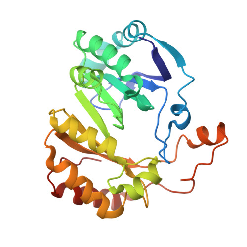
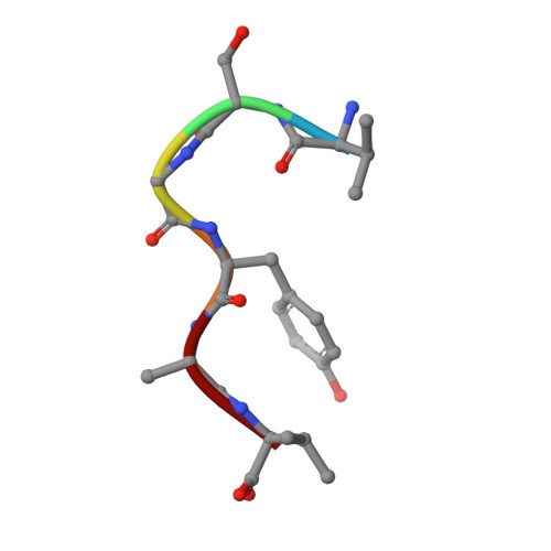
Structural basis for glycyl radical formation by pyruvate formate-lyase activating enzyme.

Vey, J.L.Yang, J.Li, M.Broderick, W.E.Broderick, J.B.Drennan, C.L.

(2008) Proc Natl Acad Sci U S A 105: 16137-16141

  • DOI: https://doi.org/10.1073/pnas.0806640105
  • Primary Citation Related Structures: 
    3C8F, 3CB8

  • PubMed Abstract: 

    Pyruvate formate-lyase activating enzyme generates a stable and catalytically essential glycyl radical on G(734) of pyruvate formate-lyase via the direct, stereospecific abstraction of a hydrogen atom from pyruvate formate-lyase. The activase performs this remarkable feat by using an iron-sulfur cluster and S-adenosylmethionine (AdoMet), thus placing it among the AdoMet radical superfamily of enzymes. We report here structures of the substrate-free and substrate-bound forms of pyruvate formate-lyase-activating enzyme, the first structures of an AdoMet radical activase. To obtain the substrate-bound structure, we have used a peptide substrate, the 7-mer RVSGYAV, which contains the sequence surrounding G(734). Our structures provide fundamental insights into the interactions between the activase and the G(734) loop of pyruvate formate-lyase and provide a structural basis for direct and stereospecific H atom abstraction from the buried G(734) of pyruvate formate-lyase.


  • Organizational Affiliation
    • Departments of Chemistry and Biology, Massachusetts Institute of Technology, Cambridge, MA 02139, USA.

Macromolecule Content 

  • Total Structure Weight: 29.67 kDa 
  • Atom Count: 1,995 
  • Modeled Residue Count: 250 
  • Deposited Residue Count: 251 
  • Unique protein chains: 2

Macromolecules

Find similar proteins by:|  3D Structure
Entity ID: 1
MoleculeChains  Sequence LengthOrganismDetailsImage
Pyruvate formate-lyase 1-activating enzyme245Escherichia coliMutation(s): 0 
Gene Names: pflA
EC: 1.97.1.4
UniProt
Find proteins for P0A9N4 (Escherichia coli (strain K12))
Explore P0A9N4 
Go to UniProtKB:  P0A9N4
Entity Groups
Sequence Clusters30% Identity50% Identity70% Identity90% Identity95% Identity100% Identity
UniProt GroupP0A9N4
Sequence Annotations
Expand
Reference Sequence
Find similar proteins by:  Sequence   |   3D Structure  
Entity ID: 2
MoleculeChains  Sequence LengthOrganismDetailsImage
peptide substrate VSGYAV6N/AMutation(s): 0 

Small Molecules

Ligands 4 Unique
IDChains Name / Formula / InChI Key2D Diagram3D Interactions
SAM

Query on SAM


Download:Ideal Coordinates CCD File
D [auth A]S-ADENOSYLMETHIONINE
C15 H22 N6 O5 S
MEFKEPWMEQBLKI-FCKMPRQPSA-N
SF4

Query on SF4


Download:Ideal Coordinates CCD File
E [auth A]IRON/SULFUR CLUSTER
Fe4 S4
LJBDFODJNLIPKO-UHFFFAOYSA-N
FMT

Query on FMT


Download:Ideal Coordinates CCD File
F [auth A],
G [auth A],
H [auth A],
I [auth A]
FORMIC ACID
C H2 O2
BDAGIHXWWSANSR-UHFFFAOYSA-N
NA

Query on NA


Download:Ideal Coordinates CCD File
C [auth A]SODIUM ION
Na
FKNQFGJONOIPTF-UHFFFAOYSA-N

Experimental Data & Validation

Experimental Data

  • Method: X-RAY DIFFRACTION
  • Resolution: 2.77 Å
  • R-Value Free:  0.254 (Depositor), 0.266 (DCC) 
  • R-Value Work:  0.229 (Depositor), 0.220 (DCC) 
  • R-Value Observed: 0.277 (Depositor) 
Space Group: P 61 2 2
Unit Cell:
Length ( Å )Angle ( ˚ )
a = 74.494α = 90
b = 74.494β = 90
c = 187.737γ = 120
Software Package:
Software NamePurpose
SOLVEphasing
CNSrefinement
PDB_EXTRACTdata extraction
Blu-Icedata collection
HKL-2000data reduction
SCALEITdata scaling

Structure Validation

View Full Validation Report


Ligand Structure Quality Assessment 


Entry History 

Deposition Data

Revision History  (Full details and data files)

  • Version 1.0: 2008-10-28
    Type: Initial release
  • Version 1.1: 2011-07-13
    Changes: Version format compliance
  • Version 1.2: 2024-02-21
    Changes: Data collection, Database references, Derived calculations