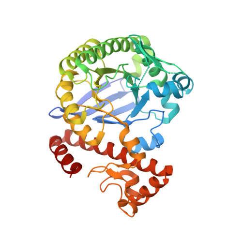
Crystal structure analysis and in silico pKa calculations suggest strong pKa shifts of ligands as driving force for high-affinity binding to TGT
Ritschel, T., Hoertner, S., Heine, A., Diederich, F., Klebe, G.(2009) Chembiochem 10: 716-727
- PubMed: 19199329 Search on PubMed
- DOI: https://doi.org/10.1002/cbic.200800782
- Primary Citation Related Structures:
2Z7K, 3C2Y - PubMed Abstract:
A novel ligand series is presented to inhibit tRNA-guanine transglycosylase (TGT), a protein with a significant role in the pathogenicity mechanism of Shigella flexneri, the causative agent of Shigellosis. The enzyme exchanges guanine in the wobble position of tRNA(Asn,Asp,His,Tyr) against a modified base. To prevent the base-exchange reaction, several series of inhibitors have already been designed, synthesized, and tested. One aim of previous studies was to address a hydrophobic pocket with different side chains attached to the parent skeletons. Disappointingly, no significant increase in binding affinity could be observed that could be explained by the disruption of a conserved water cluster. The ligand series examined in this study are based on the known scaffold lin-benzoguanine. Different side chains were introduced leading to 2-amino-lin-benzoguanines, which address a different pocket of the protein and avoid disruption of the water cluster. With the introduction of an amino group in the 2-position, a dramatic increase in binding affinity can be experienced. To explain this significant gain in binding affinity, Poisson-Boltzmann calculations were performed to explore pK(a) changes of ligand functional groups upon protein binding, they can differ significantly on going from aqueous solution to protein environment. For all complexes, a permanent protonation of the newly designed ligands is suggested, leading to a charge-assisted hydrogen bond in the protein-ligand complex. This increased strength in hydrogen bonding takes beneficial effect on binding affinity of the ligands, resulting in low-nanomolar binders. Crystal structures and docking emphasize the importance of the newly created charge-assisted hydrogen bond. A detailed analysis of the crystal structures in complex with substituted 2-amino-lin-benzoguanines indicate pronounced disorder of the attached side chains addressing the ribose 33 binding pocket. Docking suggests multiple orientations of these side chains. Obviously, an entropic advantage of the residual mobility experienced by these ligands in the bound state is beneficial and reveals an overall improved protein binding.
- Institut für Pharmazeutische Chemie, Philipps-Universität Marburg, Marbacher Weg 6, Marburg, Germany.
Organizational Affiliation: