3BZ0 | pdb_00003bz0

Lactobacillus Casei Thymidylate Synthase Ternary Complex with DUMP and the Phtalimidic Derivative C00


Experimental Data Snapshot

  • Method: X-RAY DIFFRACTION
  • Resolution: 2.70 Å
  • R-Value Free: 
    0.326 (Depositor), 0.322 (DCC) 
  • R-Value Work: 
    0.226 (Depositor), 0.226 (DCC) 
  • R-Value Observed: 
    0.235 (Depositor) 

Starting Model: experimental
View more details

wwPDB Validation 3D Report Full Report

Validation slider image for 3BZ0

Ligand Structure Quality Assessment 


This is version 1.3 of the entry. See complete history

Literature

Identification of the binding modes of N-phenylphthalimides inhibiting bacterial thymidylate synthase through X-ray crystallography screening

Mangani, S.Cancian, L.Leone, R.Pozzi, C.Lazzari, S.Luciani, R.Ferrari, S.Costi, M.P.

(2011) J Med Chem 54: 5454-5467

  • DOI: https://doi.org/10.1021/jm2005018
  • Primary Citation Related Structures: 
    3BNZ, 3BYX, 3BZ0, 3C06, 3C0A, 3IJZ, 3IK0, 3IK1

  • PubMed Abstract: 

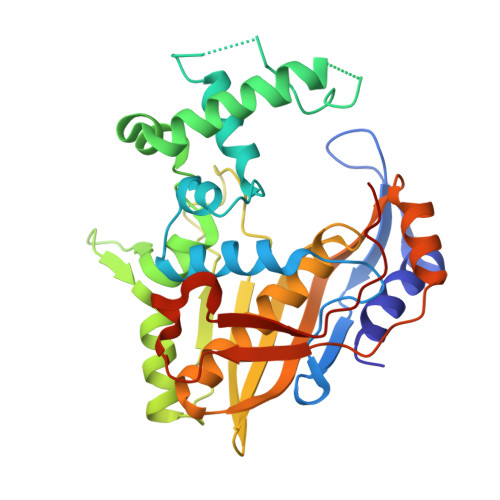
    To identify specific bacterial thymidylate synthase (TS) inhibitors, we exploited phenolphthalein (PTH), which inhibits both bacterial and human enzymes. The X-ray crystal structure of Lactobacillus casei TS (LcTS) that binds PTH showed multiple binding modes of the inhibitor, which prevented a classical structure-based drug design approach. To overcome this issue, we synthesized two phthalimidic libraries that were tested against TS enzymes and then we performed X-ray crystallographic screening of the active compounds. Compounds 6A, 8A, and 12A showed 40-fold higher affinity for bacterial TS than human TS. The X-ray crystallographic screening characterized the binding mode of six inhibitors in complexes with LcTS. Of these, 20A, 23A, and 24A showed a common unique binding mode, whereas 8A showed a different, unique binding mode. A comparative analysis of the LcTS X-ray complexes that were obtained with the pathogenic TS enabled the selection of compounds 8A and 23A as specific compounds and starting points to be exploited for the specific inhibition of pathogen enzymes.


  • Organizational Affiliation
    • Dipartimento di Scienze Farmaceutiche, Università degli Studi di Modena e Reggio Emilia, Modena, Italy. mangani@unisi.it

Macromolecule Content 

  • Total Structure Weight: 37.27 kDa 
  • Atom Count: 2,602 
  • Modeled Residue Count: 306 
  • Deposited Residue Count: 316 
  • Unique protein chains: 1

Macromolecules

Find similar proteins by:|  3D Structure
Entity ID: 1
MoleculeChains  Sequence LengthOrganismDetailsImage
Thymidylate synthase316Lacticaseibacillus caseiMutation(s): 0 
EC: 2.1.1.45
UniProt
Find proteins for P00469 (Lacticaseibacillus casei)
Explore P00469 
Go to UniProtKB:  P00469
Entity Groups
Sequence Clusters30% Identity50% Identity70% Identity90% Identity95% Identity100% Identity
UniProt GroupP00469
Sequence Annotations
Expand
Reference Sequence

Small Molecules

Ligands 2 Unique
IDChains Name / Formula / InChI Key2D Diagram3D Interactions
C00

Query on C00


Download:Ideal Coordinates CCD File
C [auth A]2-(4-hydroxybiphenyl-3-yl)-4-methyl-1H-isoindole-1,3(2H)-dione
C21 H15 N O3
ROMNEYAQUBNRLR-UHFFFAOYSA-N
UMP

Query on UMP


Download:Ideal Coordinates CCD File
B [auth A]2'-DEOXYURIDINE 5'-MONOPHOSPHATE
C9 H13 N2 O8 P
JSRLJPSBLDHEIO-SHYZEUOFSA-N

Experimental Data & Validation

Experimental Data

  • Method: X-RAY DIFFRACTION
  • Resolution: 2.70 Å
  • R-Value Free:  0.326 (Depositor), 0.322 (DCC) 
  • R-Value Work:  0.226 (Depositor), 0.226 (DCC) 
  • R-Value Observed: 0.235 (Depositor) 
Space Group: P 61 2 2
Unit Cell:
Length ( Å )Angle ( ˚ )
a = 77.916α = 90
b = 77.916β = 90
c = 224.183γ = 120
Software Package:
Software NamePurpose
REFMACrefinement
MOSFLMdata reduction
SCALAdata scaling
MOLREPphasing

Structure Validation

View Full Validation Report


Ligand Structure Quality Assessment 


Entry History 

Deposition Data

Revision History  (Full details and data files)

  • Version 1.0: 2009-01-20
    Type: Initial release
  • Version 1.1: 2011-07-13
    Changes: Version format compliance
  • Version 1.2: 2013-07-24
    Changes: Database references
  • Version 1.3: 2023-11-01
    Changes: Data collection, Database references, Derived calculations, Refinement description