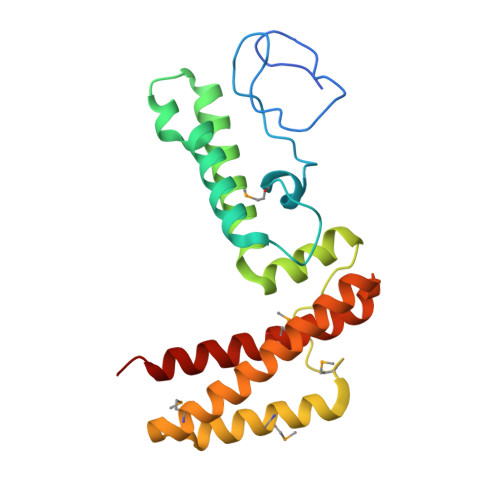
Structure of human J-type co-chaperone HscB reveals a tetracysteine metal-binding domain.
Bitto, E., Bingman, C.A., Bittova, L., Kondrashov, D.A., Bannen, R.M., Fox, B.G., Markley, J.L., Phillips, G.N.(2008) J Biological Chem 283: 30184-30192
- PubMed: 18713742
- DOI: https://doi.org/10.1074/jbc.M804746200
- Primary Citation of Related Structures:
3BVO - PubMed Abstract:
Iron-sulfur proteins play indispensable roles in a broad range of biochemical processes. The biogenesis of iron-sulfur proteins is a complex process that has become a subject of extensive research. The final step of iron-sulfur protein assembly involves transfer of an iron-sulfur cluster from a cluster-donor to a cluster-acceptor protein. This process is facilitated by a specialized chaperone system, which consists of a molecular chaperone from the Hsc70 family and a co-chaperone of the J-domain family. The 3.0 A crystal structure of a human mitochondrial J-type co-chaperone HscB revealed an L-shaped protein that resembles Escherichia coli HscB. The important difference between the two homologs is the presence of an auxiliary metal-binding domain at the N terminus of human HscB that coordinates a metal via the tetracysteine consensus motif CWXCX(9-13)FCXXCXXXQ. The domain is found in HscB homologs from animals and plants as well as in magnetotactic bacteria. The metal-binding site of the domain is structurally similar to that of rubredoxin and several zinc finger proteins containing rubredoxin-like knuckles. The normal mode analysis of HscB revealed that this L-shaped protein preferentially undergoes a scissors-like motion that correlates well with the conformational changes of human HscB observed in the crystals.
- Center for Eukaryotic Structural Genomics, University of Wisconsin-Madison, Madison, Wisconsin 53706-1544, USA.
Organizational Affiliation:


