3BVD

Structure of Surface-engineered Cytochrome ba3 Oxidase from Thermus thermophilus under Xenon Pressure, 100psi 5min


Experimental Data Snapshot

  • Method: X-RAY DIFFRACTION
  • Resolution: 3.37 Å
  • R-Value Free: 0.336 
  • R-Value Work: 0.292 

wwPDB Validation   3D Report Full Report


Ligand Structure Quality Assessment 


This is version 1.4 of the entry. See complete history


Literature

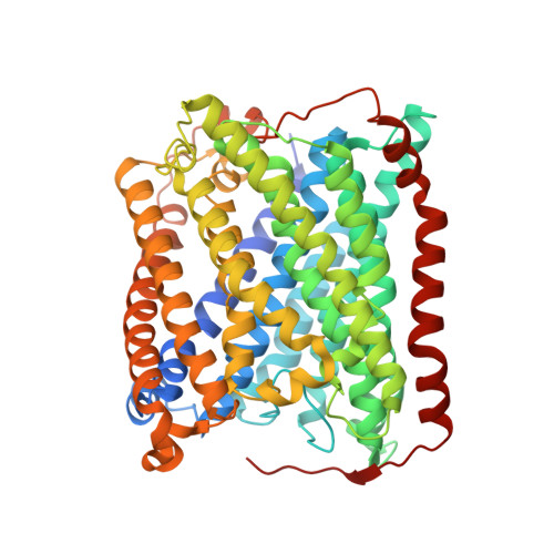
Crystallographic Studies of Xe and Kr Binding within the Large Internal Cavity of Cytochrome ba3 from Thermus thermophilus: Structural Analysis and Role of Oxygen Transport Channels in the Heme-Cu Oxidases.

Luna, V.M.Chen, Y.Fee, J.A.Stout, C.D.

(2008) Biochemistry 47: 4657-4665

  • DOI: https://doi.org/10.1021/bi800045y
  • Primary Citation of Related Structures:  
    3BVD

  • PubMed Abstract: 

    Cytochrome ba3 is a cytochrome c oxidase from the plasma membrane of Thermus thermophilus and is the preferred terminal enzyme of cellular respiration at low dioxygen tensions. Using cytochrome ba 3 crystals pressurized at varying conditions under Xe or Kr gas, and X-ray data for six crystals, we identify the relative affinities of Xe and Kr atoms for as many as seven distinct binding sites. These sites track a continuous, Y-shaped channel, 18-20 A in length, lined by hydrophobic residues, which leads from the surface of the protein where two entrance holes, representing the top of the Y, connect the bilayer to the a3-CuB center at the base of the Y. Considering the increased affinity of O2 for hydrophobic environments, the hydrophobic nature of the channel, its orientation within the bilayer, its connection to the active site, its uniform diameter, its virtually complete occupation by Xe, and its isomorphous presence in the native enzyme, we infer that the channel is a diffusion pathway for O2 into the dinuclear center of cytochrome ba3. These observations provide a basis for analyzing similar channels in other oxidases of known structure, and these structures are discussed in terms of mechanisms of O2 transport in biological systems, details of CO binding to and egress from the dinuclear center, the bifurcation of the oxygen-in and water-out pathways, and the possible role of the oxygen channel in aerobic thermophily.


  • Organizational Affiliation

    Department of Molecular Biology, The Scripps Research Institute, 10550 North Torrey Pines Road, La Jolla, CA 92037, USA.


Macromolecules
Find similar proteins by:  (by identity cutoff)  |  3D Structure
Entity ID: 1
MoleculeChains Sequence LengthOrganismDetailsImage
Cytochrome c oxidase subunit 1568Thermus thermophilusMutation(s): 1 
Gene Names: cbaA
EC: 1.9.3.1
Membrane Entity: Yes 
UniProt
Find proteins for Q5SJ79 (Thermus thermophilus (strain ATCC 27634 / DSM 579 / HB8))
Explore Q5SJ79 
Go to UniProtKB:  Q5SJ79
Entity Groups  
Sequence Clusters30% Identity50% Identity70% Identity90% Identity95% Identity100% Identity
UniProt GroupQ5SJ79
Sequence Annotations
Expand
  • Reference Sequence
Find similar proteins by:  (by identity cutoff)  |  3D Structure
Entity ID: 2
MoleculeChains Sequence LengthOrganismDetailsImage
Cytochrome c oxidase subunit 2168Thermus thermophilusMutation(s): 1 
Gene Names: cbaBctaC
EC: 1.9.3.1
Membrane Entity: Yes 
UniProt
Find proteins for Q5SJ80 (Thermus thermophilus (strain ATCC 27634 / DSM 579 / HB8))
Explore Q5SJ80 
Go to UniProtKB:  Q5SJ80
Entity Groups  
Sequence Clusters30% Identity50% Identity70% Identity90% Identity95% Identity100% Identity
UniProt GroupQ5SJ80
Sequence Annotations
Expand
  • Reference Sequence
Find similar proteins by:  (by identity cutoff)  |  3D Structure
Entity ID: 3
MoleculeChains Sequence LengthOrganismDetailsImage
Cytochrome c oxidase polypeptide 2A34Thermus thermophilusMutation(s): 0 
Gene Names: cbaD
EC: 1.9.3.1
Membrane Entity: Yes 
UniProt
Find proteins for P82543 (Thermus thermophilus (strain ATCC 27634 / DSM 579 / HB8))
Explore P82543 
Go to UniProtKB:  P82543
Entity Groups  
Sequence Clusters30% Identity50% Identity70% Identity90% Identity95% Identity100% Identity
UniProt GroupP82543
Sequence Annotations
Expand
  • Reference Sequence
Small Molecules
Ligands 5 Unique
IDChains Name / Formula / InChI Key2D Diagram3D Interactions
HAS
Query on HAS

Download Ideal Coordinates CCD File 
F [auth A]HEME-AS
C54 H64 Fe N4 O6
PDYODZVCODUKFH-ZOMLSHGTSA-L
HEM
Query on HEM

Download Ideal Coordinates CCD File 
E [auth A]PROTOPORPHYRIN IX CONTAINING FE
C34 H32 Fe N4 O4
KABFMIBPWCXCRK-RGGAHWMASA-L
XE
Query on XE

Download Ideal Coordinates CCD File 
G [auth A]
H [auth A]
I [auth A]
J [auth A]
K [auth A]
G [auth A],
H [auth A],
I [auth A],
J [auth A],
K [auth A],
L [auth A],
M [auth A]
XENON
Xe
FHNFHKCVQCLJFQ-UHFFFAOYSA-N
CUA
Query on CUA

Download Ideal Coordinates CCD File 
N [auth B]DINUCLEAR COPPER ION
Cu2
ALKZAGKDWUSJED-UHFFFAOYSA-N
CU
Query on CU

Download Ideal Coordinates CCD File 
D [auth A]COPPER (II) ION
Cu
JPVYNHNXODAKFH-UHFFFAOYSA-N
Experimental Data & Validation

Experimental Data

  • Method: X-RAY DIFFRACTION
  • Resolution: 3.37 Å
  • R-Value Free: 0.336 
  • R-Value Work: 0.292 
  • Space Group: P 41 21 2
Unit Cell:
Length ( Å )Angle ( ˚ )
a = 119.726α = 90
b = 119.726β = 90
c = 153.535γ = 90
Software Package:
Software NamePurpose
REFMACrefinement
Blu-Icedata collection
MOSFLMdata reduction
SCALAdata scaling
MOLREPphasing
REFMACphasing
CNSphasing
Cootmodel building
CNSrefinement

Structure Validation

View Full Validation Report


Ligand Structure Quality Assessment 


Entry History 

Deposition Data

Revision History  (Full details and data files)

  • Version 1.0: 2008-05-20
    Type: Initial release
  • Version 1.1: 2011-07-13
    Changes: Version format compliance
  • Version 1.2: 2017-10-25
    Changes: Refinement description
  • Version 1.3: 2021-10-20
    Changes: Database references, Derived calculations
  • Version 1.4: 2023-08-30
    Changes: Data collection, Refinement description