Probing the substrate specificity of Golgi alpha-mannosidase II by use of synthetic oligosaccharides and a catalytic nucleophile mutant.
Zhong, W., Kuntz, D.A., Ember, B., Singh, H., Moremen, K.W., Rose, D.R., Boons, G.J.(2008) J Am Chem Soc 130: 8975-8983
- PubMed: 18558690 Search on PubMedSearch on PubMed Central
- DOI: https://doi.org/10.1021/ja711248y
- Primary Citation Related Structures:
3BUB, 3BUD, 3BUI, 3BUP, 3BUQ, 3BVT, 3BVU, 3BVV, 3BVW, 3BVX - PubMed Abstract:
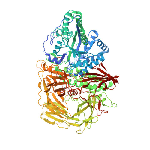
Inhibition of Golgi alpha-mannosidase II (GMII), which acts late in the N-glycan processing pathway, provides a route to blocking cancer-induced changes in cell surface oligosaccharide structures. To probe the substrate requirements of GMII, oligosaccharides were synthesized that contained an alpha(1,3)- or alpha(1,6)-linked 1-thiomannoside. Surprisingly, these oligosaccharides were not observed in X-ray crystal structures of native Drosophila GMII (dGMII). However, a mutant enzyme in which the catalytic nucleophilic aspartate was changed to alanine (D204A) allowed visualization of soaked oligosaccharides and led to the identification of the binding site for the alpha(1,3)-linked mannoside of the natural substrate. These studies also indicate that the conformational change of the bound mannoside to a high-energy B 2,5 conformation is facilitated by steric hindrance from, and the formation of strong hydrogen bonds to, Asp204. The observation that 1-thio-linked mannosides are not well tolerated by the catalytic site of dGMII led to the synthesis of a pentasaccharide containing the alpha(1,6)-linked Man of the natural substrate and the beta(1,2)-linked GlcNAc moiety proposed to be accommodated by the extended binding site of the enzyme. A cocrystal structure of this compound with the D204A enzyme revealed the molecular interactions with the beta(1,2)-linked GlcNAc. The structure is consistent with the approximately 80-fold preference of dGMII for the cleavage of substrates containing a nonreducing beta(1,2)-linked GlcNAc. By contrast, the lysosomal mannosidase lacks an equivalent GlcNAc binding site and kinetic analysis indicates oligomannoside substrates without non-reducing-terminal GlcNAc modifications are preferred, suggesting that selective inhibitors for GMII could exploit the additional binding specificity of the GlcNAc binding site.
- Complex Carbohydrate Research Center, University of Georgia, 315 Riverbend Road, Athens, Georgia 30602, USA.
Organizational Affiliation: