3BS8

Crystal structure of Glutamate 1-Semialdehyde Aminotransferase complexed with pyridoxamine-5'-phosphate From Bacillus subtilis


Experimental Data Snapshot

  • Method: X-RAY DIFFRACTION
  • Resolution: 2.30 Å
  • R-Value Free: 0.234 
  • R-Value Work: 0.193 
  • R-Value Observed: 0.195 

wwPDB Validation   3D Report Full Report


Ligand Structure Quality Assessment 


This is version 1.2 of the entry. See complete history


Literature

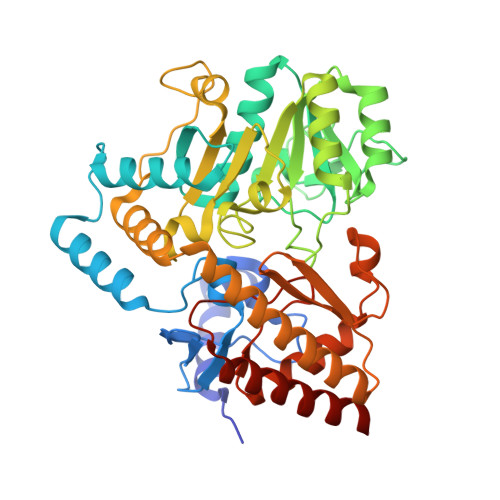
Crystal structure of Glutamate1-semialdehyde aminotransferase from Bacillus subtilis with bound pyridoxamine-5'-phosphate

Ge, H.Lv, X.Fan, J.Gao, Y.Teng, M.Niu, L.

(2010) Biochem Biophys Res Commun 402: 356-360

  • DOI: https://doi.org/10.1016/j.bbrc.2010.10.033
  • Primary Citation of Related Structures:  
    3BS8

  • PubMed Abstract: 

    Glutamate-1-semialdehyde aminotransferase (GSA-AT), also named glutamate-1-semialdehyde aminomutase (GSAM), a pyridoxamine-5'-phosphate (PMP)/pyridoxal-5'-phosphate (PLP) dependent enzyme, catalyses the transamination of the substrate glutamate-1-semialdehyde (GSA) to the product 5-Aminolevulinic acid (ALA) by an unusual intramolecular exchange of amino and oxo groups within the catalytic intermediate 4,5-diaminovalerate (DAVA). This paper presents the crystal structure of GSA-AT from Bacillus subtilis (GSA-ATBsu) in its PMP-bound form at 2.3Å resolution. The structure was determined by molecular replacement using the Synechococcus GSAM (GSAMSyn) structure as a search model. Unlike the previous reported GSAM/GSA-AT structures, GSA-ATBsu is a symmetric homodimer in the PMP-bound form, which shows the structural symmetry at the gating loop region with open state, as well as identical cofactor (PMP) binding in each monomer. This observation of PMP in combination with an "open" lid supports one characteristic feature for this enzyme, as the catalyzed reaction is believed to be initiated by PMP. Furthermore, the symmetry of GSA-ATBsu structure challenges the previously proposed negative cooperativity between monomers of this enzyme.


  • Organizational Affiliation

    Modern Experiment Technology Center, Anhui University, Hefei, Anhui 230039, China. hhge@ustc.edu


Macromolecules
Find similar proteins by:  (by identity cutoff)  |  3D Structure
Entity ID: 1
MoleculeChains Sequence LengthOrganismDetailsImage
Glutamate-1-semialdehyde 2,1-aminomutase438Bacillus subtilisMutation(s): 4 
EC: 5.4.3.8
UniProt
Find proteins for P30949 (Bacillus subtilis (strain 168))
Explore P30949 
Go to UniProtKB:  P30949
Entity Groups  
Sequence Clusters30% Identity50% Identity70% Identity90% Identity95% Identity100% Identity
UniProt GroupP30949
Sequence Annotations
Expand
  • Reference Sequence
Small Molecules
Ligands 1 Unique
IDChains Name / Formula / InChI Key2D Diagram3D Interactions
PMP
Query on PMP

Download Ideal Coordinates CCD File 
B [auth A]4'-DEOXY-4'-AMINOPYRIDOXAL-5'-PHOSPHATE
C8 H13 N2 O5 P
ZMJGSOSNSPKHNH-UHFFFAOYSA-N
Experimental Data & Validation

Experimental Data

  • Method: X-RAY DIFFRACTION
  • Resolution: 2.30 Å
  • R-Value Free: 0.234 
  • R-Value Work: 0.193 
  • R-Value Observed: 0.195 
  • Space Group: C 1 2 1
Unit Cell:
Length ( Å )Angle ( ˚ )
a = 109.251α = 90
b = 55.761β = 132.02
c = 80.504γ = 90
Software Package:
Software NamePurpose
REFMACrefinement
MAR345dtbdata collection
AUTOMARdata reduction
MOLREPphasing

Structure Validation

View Full Validation Report


Ligand Structure Quality Assessment 


Entry History 

Deposition Data

Revision History  (Full details and data files)

  • Version 1.0: 2008-12-23
    Type: Initial release
  • Version 1.1: 2011-07-13
    Changes: Version format compliance
  • Version 1.2: 2023-11-01
    Changes: Data collection, Database references, Derived calculations, Refinement description