3BS5

Crystal Structure of hCNK2-SAM/dHYP-SAM Complex


Experimental Data Snapshot

  • Method: X-RAY DIFFRACTION
  • Resolution: 2.00 Å
  • R-Value Free: 0.240 
  • R-Value Work: 0.212 
  • R-Value Observed: 0.214 

wwPDB Validation   3D Report Full Report


This is version 1.1 of the entry. See complete history


Literature

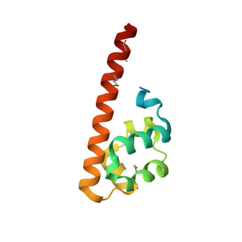
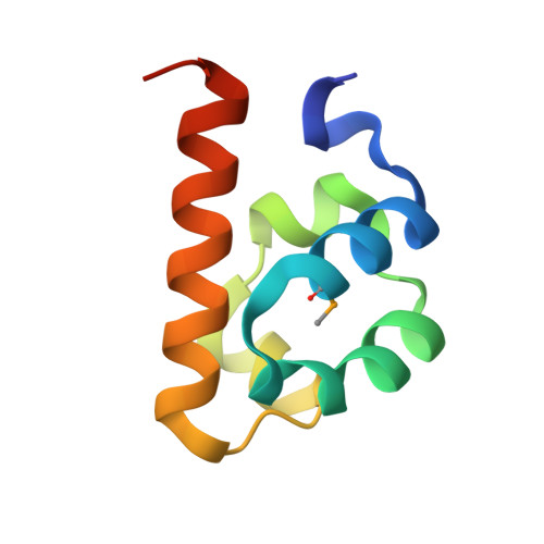
CNK and HYP form a discrete dimer by their SAM domains to mediate RAF kinase signaling.

Rajakulendran, T.Sahmi, M.Kurinov, I.Tyers, M.Therrien, M.Sicheri, F.

(2008) Proc Natl Acad Sci U S A 105: 2836-2841

  • DOI: https://doi.org/10.1073/pnas.0709705105
  • Primary Citation of Related Structures:  
    3BS5, 3BS7

  • PubMed Abstract: 

    RAF kinase functions in the mitogen-activated protein kinase (MAPK) pathway to transmit growth signals to the downstream kinases MEK and ERK. Activation of RAF catalytic activity is facilitated by a regulatory complex comprising the proteins CNK (Connector enhancer of KSR), HYP (Hyphen), and KSR (Kinase Suppressor of Ras). The sterile alpha-motif (SAM) domain found in both CNK and HYP plays an essential role in complex formation. Here, we have determined the x-ray crystal structure of the SAM domain of CNK in complex with the SAM domain of HYP. The structure reveals a single-junction SAM domain dimer of 1:1 stoichiometry in which the binding mode is a variation of polymeric SAM domain interactions. Through in vitro and in vivo mutational analyses, we show that the specific mode of dimerization revealed by the crystal structure is essential for RAF signaling and facilitates the recruitment of KSR to form the CNK/HYP/KSR regulatory complex. We present two docking-site models to account for how SAM domain dimerization might influence the formation of a higher-order CNK/HYP/KSR complex.


  • Organizational Affiliation

    Centre for Systems Biology, Samuel Lunenfeld Research Institute, Toronto, ON, Canada.


Macromolecules
Find similar proteins by:  (by identity cutoff)  |  3D Structure
Entity ID: 1
MoleculeChains Sequence LengthOrganismDetailsImage
Protein aveugle106Drosophila melanogasterMutation(s): 0 
Gene Names: ave
UniProt
Find proteins for Q8ML92 (Drosophila melanogaster)
Explore Q8ML92 
Go to UniProtKB:  Q8ML92
Entity Groups  
Sequence Clusters30% Identity50% Identity70% Identity90% Identity95% Identity100% Identity
UniProt GroupQ8ML92
Sequence Annotations
Expand
  • Reference Sequence
Find similar proteins by:  (by identity cutoff)  |  3D Structure
Entity ID: 2
MoleculeChains Sequence LengthOrganismDetailsImage
Connector enhancer of kinase suppressor of ras 280Homo sapiensMutation(s): 0 
Gene Names: CNKSR2CNK2KIAA0902KSR2
UniProt & NIH Common Fund Data Resources
Find proteins for Q8WXI2 (Homo sapiens)
Explore Q8WXI2 
Go to UniProtKB:  Q8WXI2
PHAROS:  Q8WXI2
GTEx:  ENSG00000149970 
Entity Groups  
Sequence Clusters30% Identity50% Identity70% Identity90% Identity95% Identity100% Identity
UniProt GroupQ8WXI2
Sequence Annotations
Expand
  • Reference Sequence
Small Molecules
Modified Residues  1 Unique
IDChains TypeFormula2D DiagramParent
MSE
Query on MSE
A
L-PEPTIDE LINKINGC5 H11 N O2 SeMET
Experimental Data & Validation

Experimental Data

  • Method: X-RAY DIFFRACTION
  • Resolution: 2.00 Å
  • R-Value Free: 0.240 
  • R-Value Work: 0.212 
  • R-Value Observed: 0.214 
  • Space Group: P 21 21 21
Unit Cell:
Length ( Å )Angle ( ˚ )
a = 48.477α = 90
b = 56.356β = 90
c = 84.04γ = 90
Software Package:
Software NamePurpose
SHELXphasing
REFMACrefinement
PDB_EXTRACTdata extraction
HKL-2000data collection
HKL-2000data reduction
SCALEPACKdata scaling
SHELXDphasing

Structure Validation

View Full Validation Report


Entry History 

Deposition Data

Revision History  (Full details and data files)

  • Version 1.0: 2008-02-26
    Type: Initial release
  • Version 1.1: 2011-07-13
    Changes: Version format compliance