3BOO

Structure of the C. botulinum neurotoxin serotype A with an inhibitory peptide bound


Experimental Data Snapshot

  • Method: X-RAY DIFFRACTION
  • Resolution: 1.40 Å
  • R-Value Free: 0.178 
  • R-Value Work: 0.148 
  • R-Value Observed: 0.150 

wwPDB Validation   3D Report Full Report


This is version 1.4 of the entry. See complete history


Literature

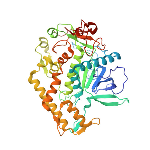
Catalytic features of the botulinum neurotoxin A light chain revealed by high resolution structure of an inhibitory peptide complex.

Silvaggi, N.R.Wilson, D.Tzipori, S.Allen, K.N.

(2008) Biochemistry 47: 5736-5745

  • DOI: https://doi.org/10.1021/bi8001067
  • Primary Citation of Related Structures:  
    3BOK, 3BON, 3BOO

  • PubMed Abstract: 

    The Clostridium botulinum neurotoxin serotype A light chain (BoNT/A-LC) is a Zn(II)-dependent metalloprotease that blocks the release of acetylcholine at the neuromuscular junction by cleaving SNAP-25, one of the SNARE proteins required for exocytosis. Because of the potential for use of the toxin in bioterrorism and the increasingly widespread application of the toxin in the medical field, there is significant interest in the development of small-molecule inhibitors of the metalloprotease. Efforts to design such inhibitors have not benefited from knowledge of how peptides bind to the active site since the enzyme-peptide structures available previously either were not occupied in the vicinity of the catalytic Zn(II) ion or did not represent the product of SNAP-25 substrate cleavage. Herein we report the 1.4 A-resolution X-ray crystal structure of a complex between the BoNT/A-LC and the inhibitory peptide N-Ac-CRATKML, the first structure of the light chain with an inhibitory peptide bound at the catalytic Zn(II) ion. The peptide is bound with the Cys S gamma atom coordinating the metal ion. Surprisingly, the cysteine sulfur is oxidized to the sulfenic acid form. Given the unstable nature of this species in solution, is it likely that oxidation occurs on the enzyme. In addition to the peptide-bound structure, we report two structures of the unliganded light chain with and without the Zn(II) cofactor bound at 1.25 and 1.20 A resolution, respectively. The two structures are nearly identical, confirming that the Zn(II) ion plays a purely catalytic role. Additionally, the structure of the Zn(II)-bound uncomplexed enzyme allows identification of the catalytic water molecule and a second water molecule that occupies the same position as the peptidic oxygen in the tetrahedral intermediate. This observation suggests that the enzyme active site is prearranged to stabilize the tetrahedral intermediate of the protease reaction.


  • Organizational Affiliation

    Department of Physiology and Biophysics, Boston University School of Medicine, Boston, Massachusetts 02118, USA.


Macromolecules
Find similar proteins by:  (by identity cutoff)  |  3D Structure
Entity ID: 1
MoleculeChains Sequence LengthOrganismDetailsImage
Neurotoxin A425Clostridium botulinumMutation(s): 0 
Gene Names: BOTAATXBNA
EC: 3.4.24.69
UniProt
Find proteins for P0DPI1 (Clostridium botulinum (strain Hall / ATCC 3502 / NCTC 13319 / Type A))
Explore P0DPI1 
Go to UniProtKB:  P0DPI1
Entity Groups  
Sequence Clusters30% Identity50% Identity70% Identity90% Identity95% Identity100% Identity
UniProt GroupP0DPI1
Sequence Annotations
Expand
  • Reference Sequence

Find similar proteins by:  Sequence   |   3D Structure  

Entity ID: 2
MoleculeChains Sequence LengthOrganismDetailsImage
N-Ac-CRATKML inhibitory peptide8N/AMutation(s): 0 
Sequence Annotations
Expand
  • Reference Sequence
Small Molecules
Ligands 1 Unique
IDChains Name / Formula / InChI Key2D Diagram3D Interactions
ZN
Query on ZN

Download Ideal Coordinates CCD File 
C [auth A]ZINC ION
Zn
PTFCDOFLOPIGGS-UHFFFAOYSA-N
Modified Residues  1 Unique
IDChains TypeFormula2D DiagramParent
CSO
Query on CSO
B
L-PEPTIDE LINKINGC3 H7 N O3 SCYS
Experimental Data & Validation

Experimental Data

  • Method: X-RAY DIFFRACTION
  • Resolution: 1.40 Å
  • R-Value Free: 0.178 
  • R-Value Work: 0.148 
  • R-Value Observed: 0.150 
  • Space Group: P 1 21 1
Unit Cell:
Length ( Å )Angle ( ˚ )
a = 50.046α = 90
b = 66.571β = 98.63
c = 65.38γ = 90
Software Package:
Software NamePurpose
DENZOdata reduction
SCALEPACKdata scaling
PHENIXrefinement
PDB_EXTRACTdata extraction
CBASSdata collection

Structure Validation

View Full Validation Report


Entry History 

Deposition Data

Revision History  (Full details and data files)

  • Version 1.0: 2008-05-20
    Type: Initial release
  • Version 1.1: 2011-07-13
    Changes: Atomic model, Database references, Derived calculations, Non-polymer description, Structure summary, Version format compliance
  • Version 1.2: 2017-10-25
    Changes: Refinement description
  • Version 1.3: 2023-08-30
    Changes: Data collection, Database references, Derived calculations, Refinement description
  • Version 1.4: 2023-11-15
    Changes: Data collection