3BOD

Structure of mouse beta-neurexin 1


Experimental Data Snapshot

  • Method: X-RAY DIFFRACTION
  • Resolution: 1.70 Å
  • R-Value Free: 0.220 
  • R-Value Work: 0.176 
  • R-Value Observed: 0.178 

wwPDB Validation   3D Report Full Report


This is version 1.3 of the entry. See complete history


Literature

Crystal Structures of beta-Neurexin 1 and beta-Neurexin 2 Ectodomains and Dynamics of Splice Insertion Sequence 4.

Koehnke, J.Jin, X.Trbovic, N.Katsamba, P.S.Brasch, J.Ahlsen, G.Scheiffele, P.Honig, B.Palmer, A.G.Shapiro, L.

(2008) Structure 16: 410-421

  • DOI: https://doi.org/10.1016/j.str.2007.12.024
  • Primary Citation of Related Structures:  
    3BOD, 3BOP

  • PubMed Abstract: 

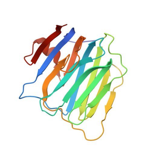
    Presynaptic neurexins (NRXs) bind to postsynaptic neuroligins (NLs) to form Ca(2+)-dependent complexes that bridge neural synapses. beta-NRXs bind NLs through their LNS domains, which contain a single site of alternative splicing (splice site 4) giving rise to two isoforms: +4 and Delta. We present crystal structures of the Delta isoforms of the LNS domains from beta-NRX1 and beta-NRX2, crystallized in the presence of Ca(2+) ions. The Ca(2+)-binding site is disordered in the beta-NRX2 structure, but the 1.7 A beta-NRX1 structure reveals a single Ca(2+) ion, approximately 12 A from the splice insertion site, with one coordinating ligand donated by a glutamic acid from an adjacent beta-NRX1 molecule. NMR studies of beta-NRX1+4 show that the insertion sequence is unstructured, and remains at least partially disordered in complex with NL. These results raise the possibility that beta-NRX insertion sequence 4 may function in roles independent of neuroligin binding.


  • Organizational Affiliation

    Department of Biochemistry and Molecular Biophysics, Columbia University, 630 West 168th Street, New York, NY 10032, USA.


Macromolecules
Find similar proteins by:  (by identity cutoff)  |  3D Structure
Entity ID: 1
MoleculeChains Sequence LengthOrganismDetailsImage
Neurexin-1-alpha178Mus musculusMutation(s): 0 
Gene Names: Nrxn1Kiaa0578
UniProt & NIH Common Fund Data Resources
Find proteins for Q9CS84 (Mus musculus)
Explore Q9CS84 
Go to UniProtKB:  Q9CS84
IMPC:  MGI:1096391
Entity Groups  
Sequence Clusters30% Identity50% Identity70% Identity90% Identity95% Identity100% Identity
UniProt GroupQ9CS84
Sequence Annotations
Expand
  • Reference Sequence
Small Molecules
Ligands 1 Unique
IDChains Name / Formula / InChI Key2D Diagram3D Interactions
CA
Query on CA

Download Ideal Coordinates CCD File 
B [auth A]CALCIUM ION
Ca
BHPQYMZQTOCNFJ-UHFFFAOYSA-N
Experimental Data & Validation

Experimental Data

  • Method: X-RAY DIFFRACTION
  • Resolution: 1.70 Å
  • R-Value Free: 0.220 
  • R-Value Work: 0.176 
  • R-Value Observed: 0.178 
  • Space Group: P 21 21 21
Unit Cell:
Length ( Å )Angle ( ˚ )
a = 44.997α = 90
b = 49.137β = 90
c = 63.556γ = 90
Software Package:
Software NamePurpose
REFMACrefinement
PDB_EXTRACTdata extraction
ADSCdata collection
DENZOdata reduction
SCALEPACKdata scaling
PHASERphasing

Structure Validation

View Full Validation Report


Entry History 

Deposition Data

Revision History  (Full details and data files)

  • Version 1.0: 2008-03-25
    Type: Initial release
  • Version 1.1: 2011-07-13
    Changes: Version format compliance
  • Version 1.2: 2017-08-02
    Changes: Refinement description, Source and taxonomy
  • Version 1.3: 2024-02-21
    Changes: Data collection, Database references, Derived calculations