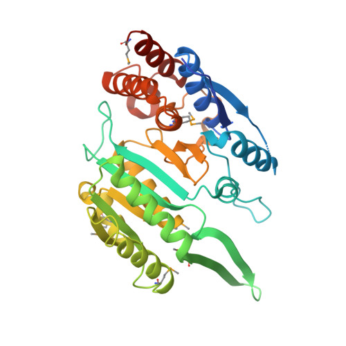
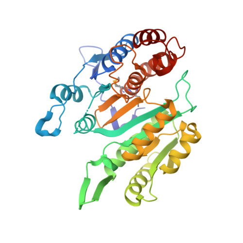
3BLV

Yeast Isocitrate Dehydrogenase with Citrate Bound in the Regulatory Subunits


Experimental Data Snapshot

  • Method: X-RAY DIFFRACTION
  • Resolution: 3.20 Å
  • R-Value Free: 0.265 
  • R-Value Work: 0.221 
  • R-Value Observed: 0.224 

Starting Model: experimental
View more details

wwPDB Validation   3D Report Full Report


This is version 1.3 of the entry. See complete history


Literature

Allosteric Motions in Structures of Yeast NAD+-specific Isocitrate Dehydrogenase.

Taylor, A.B.Hu, G.Hart, P.J.McAlister-Henn, L.

(2008) J Biol Chem 283: 10872-10880

  • DOI: https://doi.org/10.1074/jbc.M708719200
  • Primary Citation of Related Structures:  
    3BLV, 3BLW, 3BLX

  • PubMed Abstract: 

    Mitochondrial NAD(+)-specific isocitrate dehydrogenases (IDHs) are key regulators of flux through biosynthetic and oxidative pathways in response to cellular energy levels. Here we present the first structures of a eukaryotic member of this enzyme family, the allosteric, hetero-octameric, NAD(+)-specific IDH from yeast in three forms: 1) without ligands, 2) with bound analog citrate, and 3) with bound citrate + AMP. The structures reveal the molecular basis for ligand binding to homologous but distinct regulatory and catalytic sites positioned at the interfaces between IDH1 and IDH2 subunits and define pathways of communication between heterodimers and heterotetramers in the hetero-octamer. Disulfide bonds observed at the heterotetrameric interfaces in the unliganded IDH hetero-octamer are reduced in the ligand-bound forms, suggesting a redox regulatory mechanism that may be analogous to the "on-off" regulation of non-allosteric bacterial IDHs via phosphorylation. The results strongly suggest that eukaryotic IDH enzymes are exquisitely tuned to ensure that allosteric activation occurs only when concentrations of isocitrate are elevated.


  • Organizational Affiliation

    Department of Biochemistry and X-ray Crystallography Core Laboratory, The University of Texas Health Science Center, San Antonio, Texas 78229, USA.


Macromolecules
Find similar proteins by:  (by identity cutoff)  |  3D Structure
Entity ID: 1
MoleculeChains Sequence LengthOrganismDetailsImage
Isocitrate dehydrogenase [NAD] subunit 1
A, C, E, G
354Saccharomyces cerevisiaeMutation(s): 0 
Gene Names: IDH1
EC: 1.1.1.41
UniProt
Find proteins for P28834 (Saccharomyces cerevisiae (strain ATCC 204508 / S288c))
Explore P28834 
Go to UniProtKB:  P28834
Entity Groups  
Sequence Clusters30% Identity50% Identity70% Identity90% Identity95% Identity100% Identity
UniProt GroupP28834
Sequence Annotations
Expand
  • Reference Sequence
Find similar proteins by:  (by identity cutoff)  |  3D Structure
Entity ID: 2
MoleculeChains Sequence LengthOrganismDetailsImage
Isocitrate dehydrogenase [NAD] subunit 2
B, D, F, H
354Saccharomyces cerevisiaeMutation(s): 0 
Gene Names: IDH2
EC: 1.1.1.41
UniProt
Find proteins for P28241 (Saccharomyces cerevisiae (strain ATCC 204508 / S288c))
Explore P28241 
Go to UniProtKB:  P28241
Entity Groups  
Sequence Clusters30% Identity50% Identity70% Identity90% Identity95% Identity100% Identity
UniProt GroupP28241
Sequence Annotations
Expand
  • Reference Sequence
Experimental Data & Validation

Experimental Data

  • Method: X-RAY DIFFRACTION
  • Resolution: 3.20 Å
  • R-Value Free: 0.265 
  • R-Value Work: 0.221 
  • R-Value Observed: 0.224 
  • Space Group: C 1 2 1
Unit Cell:
Length ( Å )Angle ( ˚ )
a = 256.899α = 90
b = 112.949β = 106.67
c = 125.616γ = 90
Software Package:
Software NamePurpose
PHENIXrefinement
HKL-2000data reduction
HKL-2000data scaling
SHARPphasing
PHASERphasing

Structure Validation

View Full Validation Report


Entry History 

Deposition Data

Revision History  (Full details and data files)

  • Version 1.0: 2008-02-05
    Type: Initial release
  • Version 1.1: 2011-07-13
    Changes: Version format compliance
  • Version 1.2: 2023-08-30
    Changes: Data collection, Database references, Derived calculations, Refinement description
  • Version 1.3: 2023-11-15
    Changes: Data collection