3BK2

Crystal Structure Analysis of the RNase J/UMP complex


Experimental Data Snapshot

  • Method: X-RAY DIFFRACTION
  • Resolution: 2.10 Å
  • R-Value Free: 0.236 
  • R-Value Work: 0.217 
  • R-Value Observed: 0.227 

Starting Model: experimental
View more details

wwPDB Validation   3D Report Full Report


Ligand Structure Quality Assessment 


This is version 1.3 of the entry. See complete history


Literature

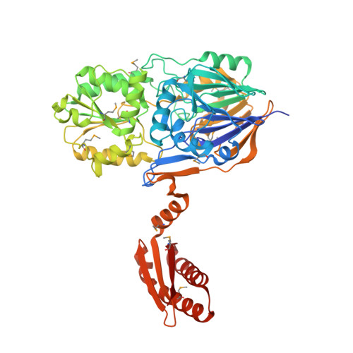
Structural insights into the dual activity of RNase J

de la Sierra-Gallay, I.L.Zig, L.Jamalli, A.Putzer, H.

(2008) Nat Struct Mol Biol 15: 206-212

  • DOI: https://doi.org/10.1038/nsmb.1376
  • Primary Citation of Related Structures:  
    3BK1, 3BK2

  • PubMed Abstract: 

    The maturation and stability of RNA transcripts is controlled by a combination of endo- and exoRNases. RNase J is unique, as it combines an RNase E-like endoribonucleolytic and a 5'-to-3' exoribonucleolytic activity in a single polypeptide. The structural basis for this dual activity is unknown. Here we report the crystal structures of Thermus thermophilus RNase J and its complex with uridine 5'-monophosphate. A binding pocket coordinating the phosphate and base moieties of the nucleotide in the vicinity of the catalytic center provide a rationale for the 5'-monophosphate-dependent 5'-to-3' exoribonucleolytic activity. We show that this dependence is strict; an initial 5'-PPP transcript cannot be degraded exonucleolytically from the 5'-end. Our results suggest that RNase J might switch promptly from endo- to exonucleolytic mode on the same RNA, a property that has important implications for RNA metabolism in numerous prokaryotic organisms and plant organelles containing RNase J orthologs.


  • Organizational Affiliation

    FRC550, Institut de Biologie Physico-Chimique, 13 rue Pierre et Marie Curie, 75005 Paris, France.


Macromolecules
Find similar proteins by:  (by identity cutoff)  |  3D Structure
Entity ID: 1
MoleculeChains Sequence LengthOrganismDetailsImage
Metal dependent hydrolase562Thermus thermophilus HB27Mutation(s): 0 
Gene Names: TTC0775
EC: 3
UniProt
Find proteins for Q72JJ7 (Thermus thermophilus (strain ATCC BAA-163 / DSM 7039 / HB27))
Explore Q72JJ7 
Go to UniProtKB:  Q72JJ7
Entity Groups  
Sequence Clusters30% Identity50% Identity70% Identity90% Identity95% Identity100% Identity
UniProt GroupQ72JJ7
Sequence Annotations
Expand
  • Reference Sequence
Small Molecules
Ligands 4 Unique
IDChains Name / Formula / InChI Key2D Diagram3D Interactions
U5P
Query on U5P

Download Ideal Coordinates CCD File 
E [auth A]URIDINE-5'-MONOPHOSPHATE
C9 H13 N2 O9 P
DJJCXFVJDGTHFX-XVFCMESISA-N
SO4
Query on SO4

Download Ideal Coordinates CCD File 
B [auth A]SULFATE ION
O4 S
QAOWNCQODCNURD-UHFFFAOYSA-L
GOL
Query on GOL

Download Ideal Coordinates CCD File 
F [auth A],
G [auth A],
H [auth A]
GLYCEROL
C3 H8 O3
PEDCQBHIVMGVHV-UHFFFAOYSA-N
ZN
Query on ZN

Download Ideal Coordinates CCD File 
C [auth A],
D [auth A]
ZINC ION
Zn
PTFCDOFLOPIGGS-UHFFFAOYSA-N
Modified Residues  1 Unique
IDChains TypeFormula2D DiagramParent
MSE
Query on MSE
A
L-PEPTIDE LINKINGC5 H11 N O2 SeMET
Experimental Data & Validation

Experimental Data

  • Method: X-RAY DIFFRACTION
  • Resolution: 2.10 Å
  • R-Value Free: 0.236 
  • R-Value Work: 0.217 
  • R-Value Observed: 0.227 
  • Space Group: F 2 2 2
Unit Cell:
Length ( Å )Angle ( ˚ )
a = 98.08α = 90
b = 118β = 90
c = 228.32γ = 90
Software Package:
Software NamePurpose
CNSrefinement
PDB_EXTRACTdata extraction
MxCuBEdata collection
XDSdata reduction
XSCALEdata scaling
MOLREPphasing

Structure Validation

View Full Validation Report


Ligand Structure Quality Assessment 


Entry History 

Deposition Data

Revision History  (Full details and data files)

  • Version 1.0: 2008-01-22
    Type: Initial release
  • Version 1.1: 2011-07-13
    Changes: Source and taxonomy, Version format compliance
  • Version 1.2: 2023-11-01
    Changes: Data collection, Database references, Derived calculations, Refinement description
  • Version 1.3: 2023-11-15
    Changes: Data collection