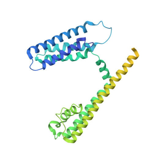
3BEH

Structure of a Bacterial Cyclic Nucleotide Regulated Ion Channel


Experimental Data Snapshot

  • Method: X-RAY DIFFRACTION
  • Resolution: 3.10 Å
  • R-Value Free: 0.286 
  • R-Value Work: 0.276 
  • R-Value Observed: 0.276 

wwPDB Validation   3D Report Full Report


Ligand Structure Quality Assessment 


This is version 1.1 of the entry. See complete history


Literature

Structure of the transmembrane regions of a bacterial cyclic nucleotide-regulated channel.

Clayton, G.M.Altieri, S.Heginbotham, L.Unger, V.M.Morais-Cabral, J.H.

(2008) Proc Natl Acad Sci U S A 105: 1511-1515

  • DOI: https://doi.org/10.1073/pnas.0711533105
  • Primary Citation of Related Structures:  
    2ZD9, 3BEH

  • PubMed Abstract: 
  • The six-transmembrane helix (6 TM) tetrameric cation channels form the largest ion channel family, some members of which are voltage-gated and others are not. There are no reported channel structures to match the wealth of functional data on the non-voltage-gated members ...

    The six-transmembrane helix (6 TM) tetrameric cation channels form the largest ion channel family, some members of which are voltage-gated and others are not. There are no reported channel structures to match the wealth of functional data on the non-voltage-gated members. We determined the structure of the transmembrane regions of the bacterial cyclic nucleotide-regulated channel MlotiK1, a non-voltage-gated 6 TM channel. The structure showed how the S1-S4 domain and its associated linker can serve as a clamp to constrain the gate of the pore and possibly function in concert with ligand-binding domains to regulate the opening of the pore. The structure also led us to hypothesize a new mechanism by which motions of the S6 inner helices can gate the ion conduction pathway at a position along the pore closer to the selectivity filter than the canonical helix bundle crossing.


    Organizational Affiliation

    Department of Molecular Biophysics and Biochemistry, Yale University, 260 Whitney Avenue, New Haven, CT 06520, USA.


Macromolecules
Find similar proteins by:  (by identity cutoff)  |  3D Structure
Entity ID: 1
MoleculeChainsSequence LengthOrganismDetailsImage
Mll3241 proteinA, B, C, D355Mesorhizobium lotiMutation(s): 0 
Membrane Entity: Yes 
UniProt
Find proteins for Q98GN8 (Mesorhizobium japonicum (strain LMG 29417 / CECT 9101 / MAFF 303099))
Explore Q98GN8 
Go to UniProtKB:  Q98GN8
Entity Groups  
Sequence Clusters30% Identity50% Identity70% Identity90% Identity95% Identity100% Identity
UniProt GroupQ98GN8
Protein Feature View
Expand
  • Reference Sequence
Experimental Data & Validation

Experimental Data

  • Method: X-RAY DIFFRACTION
  • Resolution: 3.10 Å
  • R-Value Free: 0.286 
  • R-Value Work: 0.276 
  • R-Value Observed: 0.276 
  • Space Group: H 3
Unit Cell:
Length ( Å )Angle ( ˚ )
a = 282.73α = 90
b = 282.73β = 90
c = 105.46γ = 120
Software Package:
Software NamePurpose
REFMACrefinement
XSCALEdata scaling

Structure Validation

View Full Validation Report


Ligand Structure Quality Assessment 


Entry History 

Deposition Data

Revision History  (Full details and data files)

  • Version 1.0: 2008-01-15
    Type: Initial release
  • Version 1.1: 2011-07-13
    Changes: Advisory, Version format compliance