3BDX

Crystal structure of the unstable and highly fibrillogenic Pro7Ser mutant of the Recombinant variable domain 6AJL2


Experimental Data Snapshot

  • Method: X-RAY DIFFRACTION
  • Resolution: 2.30 Å
  • R-Value Free: 0.246 
  • R-Value Work: 0.205 
  • R-Value Observed: 0.205 

wwPDB Validation   3D Report Full Report


This is version 1.3 of the entry. See complete history


Literature

A single mutation at the sheet switch region results in conformational changes favoring lambda6 light-chain fibrillogenesis.

Hernandez-Santoyo, A.del Pozo Yauner, L.Fuentes-Silva, D.Ortiz, E.Rudino-Pinera, E.Sanchez-Lopez, R.Horjales, E.Becerril, B.Rodriguez-Romero, A.

(2010) J Mol Biol 396: 280-292

  • DOI: https://doi.org/10.1016/j.jmb.2009.11.038
  • Primary Citation of Related Structures:  
    2W0K, 3B5G, 3BDX

  • PubMed Abstract: 

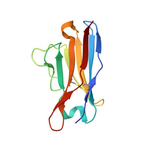
    Systemic amyloid light-chain (LC) amyloidosis is a disease process characterized by the pathological deposition of monoclonal LCs in tissue. All LC subtypes are capable of fibril formation although lambda chains, particularly those belonging to the lambda6 type, are overrepresented. Here, we report the thermodynamic and in vitro fibrillogenic properties of several mutants of the lambda6 protein 6aJL2 in which Pro7 and/or His8 was substituted by Ser or Pro. The H8P and H8S mutants were almost as stable as the wild-type protein and were poorly fibrillogenic. In contrast, the P7S mutation decreased the thermodynamic stability of 6aJL2 and greatly enhanced its capacity to form amyloid-like fibrils in vitro. The crystal structure of the P7S mutant showed that the substitution induced both local and long-distance effects, such as the rearrangement of the V(L) (variable region of the light chain)-V(L) interface. This mutant crystallized in two orthorhombic polymorphs, P2(1)2(1)2(1) and C222(1). In the latter, a monomer that was not arranged in the typical Bence-Jones dimer was observed for the first time. Crystal-packing analysis of the C222(1) lattice showed the establishment of intermolecular beta-beta interactions that involved the N-terminus and beta-strand B and that these could be relevant in the mechanism of LC fibril formation. Our results strongly suggest that Pro7 is a key residue in the conformation of the N-terminal sheet switch motif and, through long-distance interactions, is also critically involved in the contacts that stabilized the V(L) interface in lambda6 LCs.


  • Organizational Affiliation

    Instituto de Química, Universidad Nacional Autónoma de México, Circuito Exterior, Ciudad Universitaria, Mexico DF, Mexico. hersan@servidor.unam.mx


Macromolecules
Find similar proteins by:  (by identity cutoff)  |  3D Structure
Entity ID: 1
MoleculeChains Sequence LengthOrganismDetailsImage
Amyloid lambda 6 light chain variable region PIP (fragment)
A, B, C
111Homo sapiensMutation(s): 1 
Gene Names: VL gene segment 6a and JL2/3 gene segment
UniProt
Find proteins for Q96JD1 (Homo sapiens)
Explore Q96JD1 
Go to UniProtKB:  Q96JD1
Entity Groups  
Sequence Clusters30% Identity50% Identity70% Identity90% Identity95% Identity100% Identity
UniProt GroupQ96JD1
Sequence Annotations
Expand
  • Reference Sequence
Small Molecules
Ligands 3 Unique
IDChains Name / Formula / InChI Key2D Diagram3D Interactions
MES
Query on MES

Download Ideal Coordinates CCD File 
K [auth B]2-(N-MORPHOLINO)-ETHANESULFONIC ACID
C6 H13 N O4 S
SXGZJKUKBWWHRA-UHFFFAOYSA-N
GOL
Query on GOL

Download Ideal Coordinates CCD File 
G [auth A],
H [auth A],
J [auth B],
M [auth C]
GLYCEROL
C3 H8 O3
PEDCQBHIVMGVHV-UHFFFAOYSA-N
ACT
Query on ACT

Download Ideal Coordinates CCD File 
D [auth A],
E [auth A],
F [auth A],
I [auth B],
L [auth C]
ACETATE ION
C2 H3 O2
QTBSBXVTEAMEQO-UHFFFAOYSA-M
Experimental Data & Validation

Experimental Data

  • Method: X-RAY DIFFRACTION
  • Resolution: 2.30 Å
  • R-Value Free: 0.246 
  • R-Value Work: 0.205 
  • R-Value Observed: 0.205 
  • Space Group: C 2 2 21
Unit Cell:
Length ( Å )Angle ( ˚ )
a = 56.11α = 90
b = 106.12β = 90
c = 133.36γ = 90
Software Package:
Software NamePurpose
CNSrefinement
CrystalCleardata collection
MOSFLMdata reduction
SCALAdata scaling
PHASERphasing

Structure Validation

View Full Validation Report


Entry History 

Deposition Data

Revision History  (Full details and data files)

  • Version 1.0: 2008-10-28
    Type: Initial release
  • Version 1.1: 2011-07-13
    Changes: Non-polymer description, Version format compliance
  • Version 1.2: 2021-10-20
    Changes: Database references, Derived calculations
  • Version 1.3: 2023-08-30
    Changes: Data collection, Refinement description