


Crystal structure of murine interleukin-5
Patino, E., Kraich, M., Kotzsch, A., Saremba, S., Paschke, A., Sebald, W., Mueller, T.D.To be published.
Experimental Data Snapshot
Starting Model: experimental
View more details
wwPDB Validation 3D Report Full Report

Entity ID: 1 | |||||
|---|---|---|---|---|---|
| Molecule | Chains | Sequence Length | Organism | Details | Image |
| Interleukin-5 | 113 | Mus musculus | Mutation(s): 6 Gene Names: Il5 | 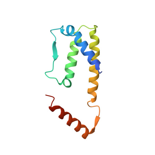 | |
UniProt | |||||
Entity Groups | |||||
| Sequence Clusters | 30% Identity50% Identity70% Identity90% Identity95% Identity100% Identity | ||||
| UniProt Group | P04401 | ||||
Sequence AnnotationsExpand | |||||
Reference Sequence | |||||
| Length ( Å ) | Angle ( ˚ ) |
|---|---|
| a = 39.193 | α = 90 |
| b = 47.085 | β = 96.41 |
| c = 55.053 | γ = 90 |
| Software Name | Purpose |
|---|---|
| REFMAC | refinement |
| CrystalClear | data collection |
| CrystalClear | data reduction |
| CrystalClear | data scaling |
| PHASER | phasing |


