Characterization of the N-Acetyl-5-neuraminic Acid-binding Site of the Extracytoplasmic Solute Receptor (SiaP) of Nontypeable Haemophilus influenzae Strain 2019
Johnston, J.W., Coussens, N.P., Allen, S., Houtman, J.C., Turner, K.H., Zaleski, A., Ramaswamy, S., Gibson, B.W., Apicella, M.A.(2008) J Biological Chem 283: 855-865
- PubMed: 17947229 Search on PubMed
- DOI: https://doi.org/10.1074/jbc.M706603200
- Primary Citation Related Structures:
3B50 - PubMed Abstract:
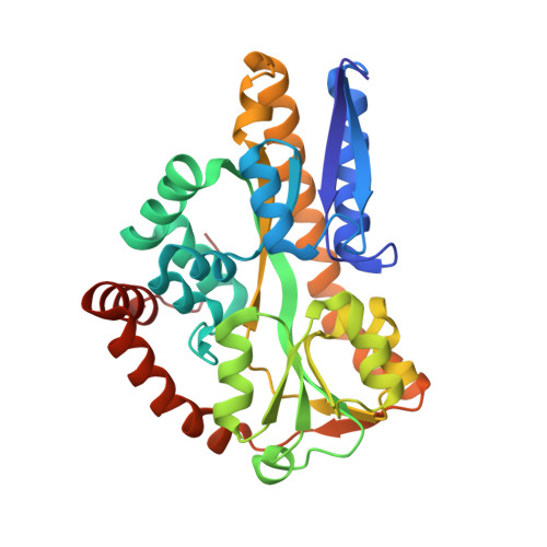
Nontypeable Haemophilus influenzae is an opportunistic human pathogen causing otitis media in children and chronic bronchitis and pneumonia in patients with chronic obstructive pulmonary disease. The outer membrane of nontypeable H. influenzae is dominated by lipooligosaccharides (LOS), many of which incorporate sialic acid as a terminal nonreducing sugar. Sialic acid has been demonstrated to be an important factor in the survival of the bacteria within the host environment. H. influenzae is incapable of synthesizing sialic acid and is dependent on scavenging free sialic acid from the host environment. To achieve this, H. influenzae utilizes a tripartite ATP-independent periplasmic transporter. In this study, we characterize the binding site of the extracytoplasmic solute receptor (SiaP) from nontypeable H. influenzae strain 2019. A crystal structure of N-acetyl-5-neuraminic acid (Neu5Ac)-bound SiaP was determined to 1.4A resolution. Thermodynamic characterization of Neu5Ac binding shows this interaction is enthalpically driven with a substantial unfavorable contribution from entropy. This is expected because the binding of SiaP to Neu5Ac is mediated by numerous hydrogen bonds and has several buried water molecules. Point mutations targeting specific amino acids were introduced in the putative binding site. Complementation with the mutated siaP constructs resulted either in full, partial, or no complementation, depending on the role of specific residues. Mass spectrometry analysis of the O-deacylated LOS of the R127K point mutation confirmed the observation of reduced incorporation of Neu5Ac into the LOS. The decreased ability of H. influenzae to import sialic acid had negative effects on resistance to complement-mediated killing and viability of biofilms in vitro, confirming the importance of sialic acid transport to the bacterium.
- Department of Microbiology, University of Iowa, Iowa City, Iowa 52242, USA.
Organizational Affiliation: