Mapping of protein-protein interaction sites in the plant-type [2Fe-2S] ferredoxin.
Kameda, H., Hirabayashi, K., Wada, K., Fukuyama, K.(2011) PLoS One 6: e21947-e21947
- PubMed: 21760931 Search on PubMedSearch on PubMed Central
- DOI: https://doi.org/10.1371/journal.pone.0021947
- Primary Citation Related Structures:
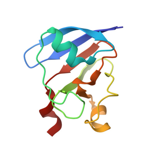
3AV8 - PubMed Abstract:
Knowing the manner of protein-protein interactions is vital for understanding biological events. The plant-type [2Fe-2S] ferredoxin (Fd), a well-known small iron-sulfur protein with low redox potential, partitions electrons to a variety of Fd-dependent enzymes via specific protein-protein interactions. Here we have refined the crystal structure of a recombinant plant-type Fd I from the blue green alga Aphanothece sacrum (AsFd-I) at 1.46 Å resolution on the basis of the synchrotron radiation data. Incorporating the revised amino-acid sequence, our analysis corrects the 3D structure previously reported; we identified the short α-helix (67-71) near the active center, which is conserved in other plant-type [2Fe-2S] Fds. Although the 3D structures of the four molecules in the asymmetric unit are similar to each other, detailed comparison of the four structures revealed the segments whose conformations are variable. Structural comparison between the Fds from different sources showed that the distribution of the variable segments in AsFd-I is highly conserved in other Fds, suggesting the presence of intrinsically flexible regions in the plant-type [2Fe-2S] Fd. A few structures of the complexes with Fd-dependent enzymes clearly demonstrate that the protein-protein interactions are achieved through these variable regions in Fd. The results described here will provide a guide for interpreting the biochemical and mutational studies that aim at the manner of interactions with Fd-dependent enzymes.
- Department of Biological Sciences, Graduate School of Science, Osaka University, Toyonaka, Osaka, Japan.
Organizational Affiliation: