3ATE | pdb_00003ate

Crystal Structure of the Kir3.2 Cytoplasmic Domain (Na+-free crystal soaked in 10 mM praseodymium (III) acetate)


Experimental Data Snapshot

  • Method: X-RAY DIFFRACTION
  • Resolution: 3.20 Å
  • R-Value Free: 
    0.325 (Depositor), 0.325 (DCC) 
  • R-Value Work: 
    0.267 (Depositor), 0.257 (DCC) 
  • R-Value Observed: 
    0.274 (Depositor) 

wwPDB Validation 3D Report Full Report

Validation slider image for 3ATE

This is version 1.4 of the entry. See complete history

Literature

Interactions of cations with the cytoplasmic pores of inward rectifier K(+) channels in the closed state

Inanobe, A.Nakagawa, A.Kurachi, Y.

(2011) J Biological Chem 286: 41801-41811

  • DOI: https://doi.org/10.1074/jbc.M111.278531
  • Primary Citation Related Structures: 
    3AT8, 3AT9, 3ATA, 3ATB, 3ATD, 3ATE, 3ATF

  • PubMed Abstract: 

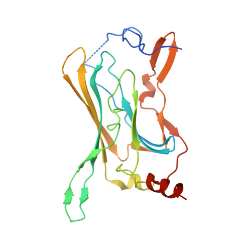
    Ion channels gate at membrane-embedded domains by changing their conformation along the ion conduction pathway. Inward rectifier K(+) (Kir) channels possess a unique extramembrane cytoplasmic domain that extends this pathway. However, the relevance and contribution of this domain to ion permeation remain unclear. By qualitative x-ray crystallographic analysis, we found that the pore in the cytoplasmic domain of Kir3.2 binds cations in a valency-dependent manner and does not allow the displacement of Mg(2+) by monovalent cations or spermine. Electrophysiological analyses revealed that the cytoplasmic pore of Kir3.2 selectively binds positively charged molecules and has a higher affinity for Mg(2+) when it has a low probability of being open. The selective blocking of chemical modification of the side chain of pore-facing residues by Mg(2+) indicates that the mode of binding of Mg(2+) is likely to be similar to that observed in the crystal structure. These results indicate that the Kir3.2 crystal structure has a closed conformation with a negative electrostatic field potential at the cytoplasmic pore, the potential of which may be controlled by conformational changes in the cytoplasmic domain to regulate ion diffusion along the pore.


  • Organizational Affiliation
    • Department of Pharmacology, Graduate School of Medicine, Osaka University, Osaka 565-0871, Japan; Center for Advanced Medical Engineering and Informatics, Osaka University, Osaka 565-0871, Japan. Electronic address: inanobe@pharma2.med.osaka-u.ac.jp.

Macromolecule Content 

  • Total Structure Weight: 24.17 kDa 
  • Atom Count: 1,576 
  • Modeled Residue Count: 196 
  • Deposited Residue Count: 208 
  • Unique protein chains: 1

Macromolecules

Find similar proteins by:|  3D Structure
Entity ID: 1
MoleculeChains  Sequence LengthOrganismDetailsImage
Potassium inwardly-rectifying channel, subfamily J, member 6208Mus musculusMutation(s): 0 
UniProt & NIH Common Fund Data Resources
Find proteins for P48542 (Mus musculus)
Explore P48542 
Go to UniProtKB:  P48542
IMPC:  MGI:104781
Entity Groups
Sequence Clusters30% Identity50% Identity70% Identity90% Identity95% Identity100% Identity
UniProt GroupP48542
Sequence Annotations
Expand
Reference Sequence

Experimental Data & Validation

Experimental Data

  • Method: X-RAY DIFFRACTION
  • Resolution: 3.20 Å
  • R-Value Free:  0.325 (Depositor), 0.325 (DCC) 
  • R-Value Work:  0.267 (Depositor), 0.257 (DCC) 
  • R-Value Observed: 0.274 (Depositor) 
Space Group: I 4 2 2
Unit Cell:
Length ( Å )Angle ( ˚ )
a = 82.775α = 90
b = 82.775β = 90
c = 172.894γ = 90
Software Package:
Software NamePurpose
SCALEPACKdata scaling
MOLREPphasing
REFMACrefinement
PDB_EXTRACTdata extraction
DENZOdata reduction

Structure Validation

View Full Validation Report


Entry History 

Deposition Data

Revision History  (Full details and data files)

  • Version 1.0: 2011-10-19
    Type: Initial release
  • Version 1.1: 2013-06-05
    Changes: Database references
  • Version 1.2: 2017-08-16
    Changes: Advisory, Refinement description, Source and taxonomy
  • Version 1.3: 2017-10-11
    Changes: Refinement description
  • Version 1.4: 2024-03-13
    Changes: Advisory, Data collection, Database references, Derived calculations