3ASM | pdb_00003asm

Crystal structure of Q54A mutant protein of Bst-RNase HIII


Experimental Data Snapshot

  • Method: X-RAY DIFFRACTION
  • Resolution: 2.60 Å
  • R-Value Free: 
    0.233 (Depositor), 0.228 (DCC) 
  • R-Value Work: 
    0.204 (Depositor), 0.199 (DCC) 
  • R-Value Observed: 
    0.205 (Depositor) 

Starting Model: experimental
View more details

wwPDB Validation 3D Report Full Report

Validation slider image for 3ASM

This is version 1.3 of the entry. See complete history

Literature

Identification of the substrate binding site in the N-terminal TBP-like domain of RNase H3.

Miyashita, S.Tadokoro, T.Angkawidjaja, C.You, D.-J.Koga, Y.Takano, K.Kanaya, S.

(2011) FEBS Lett 585: 2313-2317

  • DOI: https://doi.org/10.1016/j.febslet.2011.05.064
  • Primary Citation Related Structures: 
    3ASM

  • PubMed Abstract: 

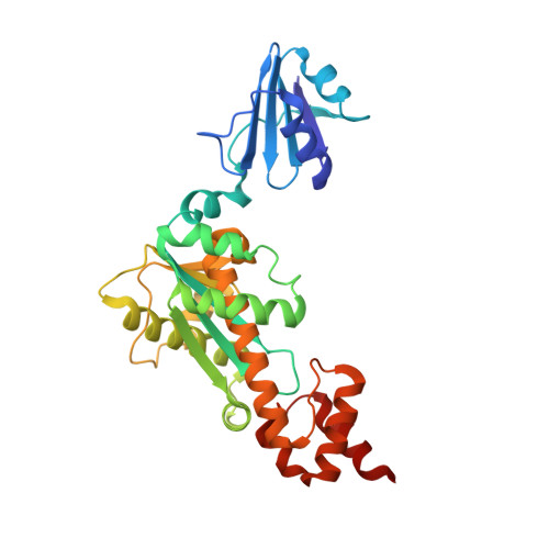
    Ribonuclease H3 from Bacillus stearothermophilus (Bst-RNase H3) has the N-terminal TBP-like substrate-binding domain. To identify the substrate binding site in this domain, the mutant proteins of the intact protein and isolated N-domain, in which six of the seventeen residues corresponding to those involved in DNA binding of TBP are individually mutated to Ala, were constructed. All of them exhibited decreased enzymatic activities and/or substrate-binding affinities when compared to those of the parent proteins, suggesting that the N-terminal domain of RNase H3 uses the flat surface of the β-sheet for substrate binding as TBP to bind DNA. This domain may greatly change conformation upon substrate binding.


  • Organizational Affiliation
    • Department of Material and Life Science, Graduate School of Engineering, Osaka University, Suita, Osaka, Japan.

Macromolecule Content 

  • Total Structure Weight: 33.82 kDa 
  • Atom Count: 2,417 
  • Modeled Residue Count: 308 
  • Deposited Residue Count: 310 
  • Unique protein chains: 1

Macromolecules

Find similar proteins by:|  3D Structure
Entity ID: 1
MoleculeChains  Sequence LengthOrganismDetailsImage
Ribonuclease HIII310Geobacillus stearothermophilusMutation(s): 1 
EC: 3.1.26.4
UniProt
Find proteins for Q6L6Q4 (Geobacillus stearothermophilus)
Explore Q6L6Q4 
Go to UniProtKB:  Q6L6Q4
Entity Groups
Sequence Clusters30% Identity50% Identity70% Identity90% Identity95% Identity100% Identity
UniProt GroupQ6L6Q4
Sequence Annotations
Expand
Reference Sequence

Experimental Data & Validation

Experimental Data

  • Method: X-RAY DIFFRACTION
  • Resolution: 2.60 Å
  • R-Value Free:  0.233 (Depositor), 0.228 (DCC) 
  • R-Value Work:  0.204 (Depositor), 0.199 (DCC) 
  • R-Value Observed: 0.205 (Depositor) 
Space Group: P 21 21 2
Unit Cell:
Length ( Å )Angle ( ˚ )
a = 67.249α = 90
b = 109.106β = 90
c = 48.329γ = 90
Software Package:
Software NamePurpose
BL38data collection
MOLREPphasing
REFMACrefinement
HKL-2000data reduction
HKL-2000data scaling

Structure Validation

View Full Validation Report


Entry History 

Deposition Data

Revision History  (Full details and data files)

  • Version 1.0: 2011-06-08
    Type: Initial release
  • Version 1.1: 2011-07-13
    Changes: Version format compliance
  • Version 1.2: 2011-11-23
    Changes: Database references
  • Version 1.3: 2023-11-01
    Changes: Data collection, Database references, Refinement description