3AO0 | pdb_00003ao0

Crystal structure of ethanolamine ammonia-lyase from Escherichia coli complexed with CN-CBL and (S)-2-amino-1-propanol


Experimental Data Snapshot

  • Method: X-RAY DIFFRACTION
  • Resolution: 2.25 Å
  • R-Value Free: 
    0.257 (Depositor), 0.273 (DCC) 
  • R-Value Work: 
    0.235 (Depositor), 0.246 (DCC) 
  • R-Value Observed: 
    0.236 (Depositor) 

Starting Model: experimental
View more details

wwPDB Validation 3D Report Full Report

Validation slider image for 3AO0

Ligand Structure Quality Assessment 


This is version 2.0 of the entry. See complete history

Literature

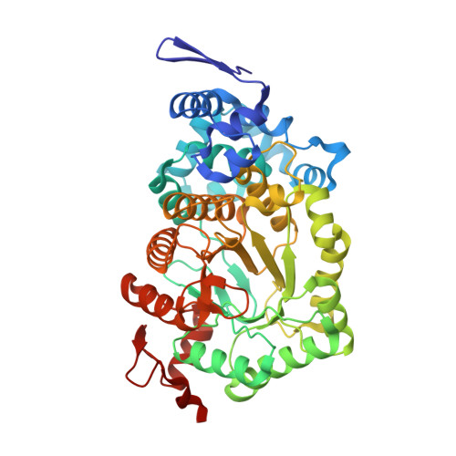
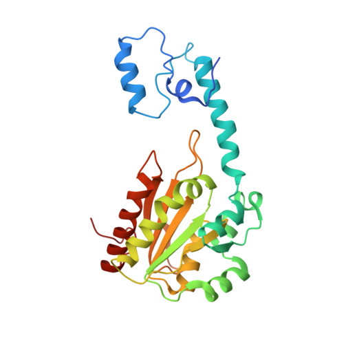
How coenzyme B12-dependent ethanolamine ammonia-lyase deals with both enantiomers of 2-amino-1-propanol as substrates: structure-based rationalization.

Shibata, N.Higuchi, Y.Toraya, T.

(2011) Biochemistry 50: 591-598

  • DOI: https://doi.org/10.1021/bi101696h
  • Primary Citation Related Structures: 
    3ANY, 3AO0

  • PubMed Abstract: 

    Coenzyme B(12)-dependent ethanolamine ammonia-lyase acts on both enantiomers of the substrate 2-amino-1-propanol [Diziol, P., et al. (1980) Eur. J. Biochem. 106, 211-224]. To rationalize this apparent lack of stereospecificity and the enantiomer-specific stereochemical courses of the deamination, we analyzed the X-ray structures of enantiomer-bound forms of the enzyme-cyanocobalamin complex. The lower affinity for the (R)-enantiomer may be due to the conformational change of the Valα326 side chain of the enzyme. In a manner consistent with the reported experimental results, we can predict that the pro-S hydrogen atom on C1 is abstracted by the adenosyl radical from both enantiomeric substrates, because it is the nearest one in both enantiomer-bound forms. We also predicted that the NH(2) group migrates from C2 to C1 by a suprafacial shift, with inversion of configuration at C1 for both enantiomeric substrates, although the absolute configuration of the 1-amino-1-propanol intermediate is not yet known. Reported labeling experiments demonstrate that (R)-2-amino-1-propanol is deaminated by the enzyme with inversion of configuration at C2, whereas the (S)-enantiomer is deaminated with retention. By taking these results into consideration, we can predict the rotameric radical intermediate from the (S)-enantiomer undergoes flipping to the rotamer from the (R)-enantiomer before the hydrogen back-abstraction. This suggests the preference of the enzyme active site for the rotamer from the (R)-enantiomer in equilibration. This preference might be explained in terms of the steric repulsion of the (S)-enantiomer-derived product radical at C3 with the Pheα329 and Leuα402 residues.


  • Organizational Affiliation
    • Department of Life Science, Graduate School of Life Science, University of Hyogo, 3-2-1 Koto, Kamigori-cho, Ako-gun, Hyogo, Japan. shibach@sci.u-hyogo.ac.jp

Macromolecule Content 

  • Total Structure Weight: 159.27 kDa 
  • Atom Count: 11,550 
  • Modeled Residue Count: 1,410 
  • Deposited Residue Count: 1,432 
  • Unique protein chains: 2

Macromolecules

Find similar proteins by:|  3D Structure
Entity ID: 1
MoleculeChains  Sequence LengthOrganismDetailsImage
Ethanolamine ammonia-lyase heavy chain
A, C
453Escherichia coli K-12Mutation(s): 0 
Gene Names: eutBb2441JW2434
EC: 4.3.1.7
UniProt
Find proteins for P0AEJ6 (Escherichia coli (strain K12))
Explore P0AEJ6 
Go to UniProtKB:  P0AEJ6
Entity Groups
Sequence Clusters30% Identity50% Identity70% Identity90% Identity95% Identity100% Identity
UniProt GroupP0AEJ6
Sequence Annotations
Expand
Reference Sequence
Find similar proteins by:|  3D Structure
Entity ID: 2
MoleculeChains  Sequence LengthOrganismDetailsImage
Ethanolamine ammonia-lyase light chain
B, D
263Escherichia coli K-12Mutation(s): 0 
Gene Names: eutCb2440JW2433
EC: 4.3.1.7
UniProt
Find proteins for P19636 (Escherichia coli (strain K12))
Explore P19636 
Go to UniProtKB:  P19636
Entity Groups
Sequence Clusters30% Identity50% Identity70% Identity90% Identity95% Identity100% Identity
UniProt GroupP19636
Sequence Annotations
Expand
Reference Sequence

Experimental Data & Validation

Experimental Data

  • Method: X-RAY DIFFRACTION
  • Resolution: 2.25 Å
  • R-Value Free:  0.257 (Depositor), 0.273 (DCC) 
  • R-Value Work:  0.235 (Depositor), 0.246 (DCC) 
  • R-Value Observed: 0.236 (Depositor) 
Space Group: P 63
Unit Cell:
Length ( Å )Angle ( ˚ )
a = 243.85α = 90
b = 243.85β = 90
c = 76.806γ = 120
Software Package:
Software NamePurpose
BSSdata collection
REFMACrefinement
HKL-2000data reduction
HKL-2000data scaling

Structure Validation

View Full Validation Report


Ligand Structure Quality Assessment 


Entry History 

Deposition Data

  • Released Date: 2011-08-03 
  • Deposition Author(s): Shibata, N.

Revision History  (Full details and data files)

  • Version 1.0: 2011-08-03
    Type: Initial release
  • Version 1.1: 2012-10-24
    Changes: Non-polymer description
  • Version 1.2: 2023-11-01
    Changes: Data collection, Database references, Derived calculations, Refinement description
  • Version 2.0: 2023-11-15
    Changes: Atomic model, Data collection