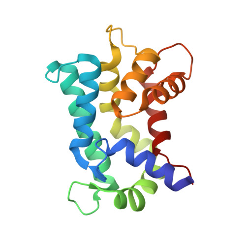
3AKB

Structural basis for prokaryotic calcium-mediated regulation by a Streptomyces coelicolor calcium-binding protein


Experimental Data Snapshot

  • Method: X-RAY DIFFRACTION
  • Resolution: 1.50 Å
  • R-Value Free: 0.232 
  • R-Value Work: 0.203 
  • R-Value Observed: 0.205 

wwPDB Validation   3D Report Full Report


This is version 1.2 of the entry. See complete history


Literature

Structural basis for prokaryotic calciummediated regulation by a Streptomyces coelicolor calcium binding protein

Zhao, X.Pang, H.Wang, S.Zhou, W.Yang, K.Bartlam, M.

(2010) Protein Cell 1: 771-779

  • DOI: https://doi.org/10.1007/s13238-010-0085-z
  • Primary Citation of Related Structures:  
    3AKA, 3AKB

  • PubMed Abstract: 

    The important and diverse regulatory roles of Ca(2+) in eukaryotes are conveyed by the EF-hand containing calmodulin superfamily. However, the calcium-regulatory proteins in prokaryotes are still poorly understood. In this study, we report the three-dimensional structure of the calcium-binding protein from Streptomyces coelicolor, named CabD, which shares low sequence homology with other known helix-loop-helix EF-hand proteins. The CabD structure should provide insights into the biological role of the prokaryotic calcium-binding proteins. The unusual structural features of CabD compared with prokaryotic EF-hand proteins and eukaryotic sarcoplasmic calcium-binding proteins, including the bending conformation of the first C-terminal α-helix, unpaired ligand-binding EF-hands and the lack of the extreme C-terminal loop region, suggest it may have a distinct and significant function in calcium-mediated bacterial physiological processes, and provide a structural basis for potential calcium-mediated regulatory roles in prokaryotes.


  • Organizational Affiliation

    Laboratory of Structural Biology, Tsinghua University, Beijing 100084, China.


Macromolecules
Find similar proteins by:  (by identity cutoff)  |  3D Structure
Entity ID: 1
MoleculeChains Sequence LengthOrganismDetailsImage
Putative calcium binding protein166Streptomyces coelicolor A3(2)Mutation(s): 2 
Gene Names: cabdSC6F11.09SCO4411
UniProt
Find proteins for Q9F377 (Streptomyces coelicolor (strain ATCC BAA-471 / A3(2) / M145))
Explore Q9F377 
Go to UniProtKB:  Q9F377
Entity Groups  
Sequence Clusters30% Identity50% Identity70% Identity90% Identity95% Identity100% Identity
UniProt GroupQ9F377
Sequence Annotations
Expand
  • Reference Sequence
Small Molecules
Ligands 1 Unique
IDChains Name / Formula / InChI Key2D Diagram3D Interactions
CA
Query on CA

Download Ideal Coordinates CCD File 
B [auth A]
C [auth A]
D [auth A]
E [auth A]
F [auth A]
B [auth A],
C [auth A],
D [auth A],
E [auth A],
F [auth A],
G [auth A],
H [auth A],
I [auth A],
J [auth A],
K [auth A],
L [auth A]
CALCIUM ION
Ca
BHPQYMZQTOCNFJ-UHFFFAOYSA-N
Experimental Data & Validation

Experimental Data

  • Method: X-RAY DIFFRACTION
  • Resolution: 1.50 Å
  • R-Value Free: 0.232 
  • R-Value Work: 0.203 
  • R-Value Observed: 0.205 
  • Space Group: P 21 21 21
Unit Cell:
Length ( Å )Angle ( ˚ )
a = 33.144α = 90
b = 51.137β = 90
c = 87.26γ = 90
Software Package:
Software NamePurpose
MAR345dtbdata collection
SOLVEphasing
REFMACrefinement
HKL-2000data reduction
HKL-2000data scaling

Structure Validation

View Full Validation Report


Entry History 

Deposition Data

Revision History  (Full details and data files)

  • Version 1.0: 2011-01-26
    Type: Initial release
  • Version 1.1: 2011-07-13
    Changes: Version format compliance
  • Version 1.2: 2024-03-13
    Changes: Data collection, Database references, Derived calculations