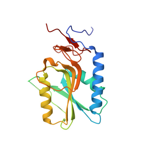
3ACC

Crystal structure of hypoxanthine-guanine phosphoribosyltransferase with GMP from Thermus thermophilus HB8


Experimental Data Snapshot

  • Method: X-RAY DIFFRACTION
  • Resolution: 2.16 Å
  • R-Value Free: 0.250 
  • R-Value Work: 0.210 
  • R-Value Observed: 0.210 

Starting Model: experimental
View more details

wwPDB Validation   3D Report Full Report


Ligand Structure Quality Assessment 


This is version 1.2 of the entry. See complete history


Literature

Structures of hypoxanthine-guanine phosphoribosyltransferase (TTHA0220) from Thermus thermophilus HB8.

Kanagawa, M.Baba, S.Ebihara, A.Shinkai, A.Hirotsu, K.Mega, R.Kim, K.Kuramitsu, S.Sampei, G.Kawai, G.

(2010) Acta Crystallogr Sect F Struct Biol Cryst Commun 66: 893-898

  • DOI: https://doi.org/10.1107/S1744309110023079
  • Primary Citation of Related Structures:  
    3ACB, 3ACC, 3ACD

  • PubMed Abstract: 

    Hypoxanthine-guanine phosphoribosyltransferase (HGPRTase), which is a key enzyme in the purine-salvage pathway, catalyzes the synthesis of IMP or GMP from alpha-D-phosphoribosyl-1-pyrophosphate and hypoxanthine or guanine, respectively. Structures of HGPRTase from Thermus thermophilus HB8 in the unliganded form, in complex with IMP and in complex with GMP have been determined at 2.1, 1.9 and 2.2 A resolution, respectively. The overall fold of the IMP complex was similar to that of the unliganded form, but the main-chain and side-chain atoms of the active site moved to accommodate IMP. The overall folds of the IMP and GMP complexes were almost identical to each other. Structural comparison of the T. thermophilus HB8 enzyme with 6-oxopurine PRTases for which structures have been determined showed that these enzymes can be tentatively divided into groups I and II and that the T. thermophilus HB8 enzyme belongs to group I. The group II enzymes are characterized by an N-terminal extension with additional secondary elements and a long loop connecting the second alpha-helix and beta-strand compared with the group I enzymes.


  • Organizational Affiliation

    RIKEN SPring-8 Center, Harima Institute, 1-1-1 Kouto, Sayo, Hyogo 679-5148, Japan.


Macromolecules
Find similar proteins by:  (by identity cutoff)  |  3D Structure
Entity ID: 1
MoleculeChains Sequence LengthOrganismDetailsImage
Hypoxanthine-guanine phosphoribosyltransferase181Thermus thermophilus HB8Mutation(s): 0 
Gene Names: TTHA0220
EC: 2.4.2.8
UniProt
Find proteins for Q5SLS3 (Thermus thermophilus (strain ATCC 27634 / DSM 579 / HB8))
Explore Q5SLS3 
Go to UniProtKB:  Q5SLS3
Entity Groups  
Sequence Clusters30% Identity50% Identity70% Identity90% Identity95% Identity100% Identity
UniProt GroupQ5SLS3
Sequence Annotations
Expand
  • Reference Sequence
Experimental Data & Validation

Experimental Data

  • Method: X-RAY DIFFRACTION
  • Resolution: 2.16 Å
  • R-Value Free: 0.250 
  • R-Value Work: 0.210 
  • R-Value Observed: 0.210 
  • Space Group: P 65 2 2
Unit Cell:
Length ( Å )Angle ( ˚ )
a = 67.139α = 90
b = 67.139β = 90
c = 152.543γ = 120
Software Package:
Software NamePurpose
HKL-2000data collection
MOLREPphasing
CNSrefinement
HKL-2000data reduction
HKL-2000data scaling

Structure Validation

View Full Validation Report


Ligand Structure Quality Assessment 


Entry History 

Deposition Data

Revision History  (Full details and data files)

  • Version 1.0: 2010-02-09
    Type: Initial release
  • Version 1.1: 2011-07-13
    Changes: Version format compliance
  • Version 1.2: 2023-11-01
    Changes: Data collection, Database references, Derived calculations, Refinement description