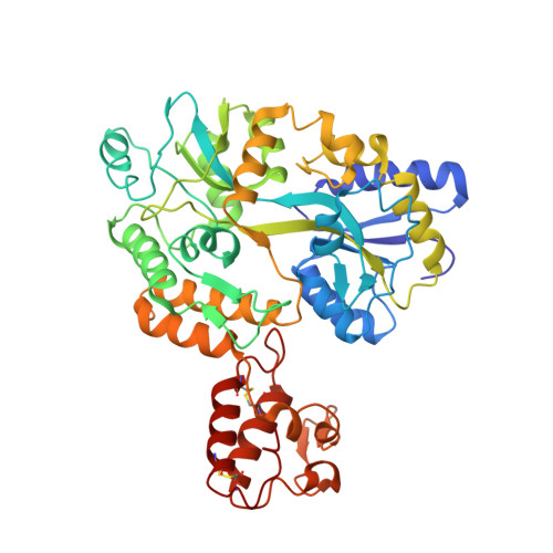
3A3C

Crystal structure of TIM40/MIA40 fusing MBP, C296S and C298S mutant


Experimental Data Snapshot

  • Method: X-RAY DIFFRACTION
  • Resolution: 2.50 Å
  • R-Value Free: 0.316 
  • R-Value Work: 0.254 
  • R-Value Observed: 0.254 

Starting Model: experimental
View more details

wwPDB Validation   3D Report Full Report


This is version 2.2 of the entry. See complete history


Literature

Structural basis of yeast Tim40/Mia40 as an oxidative translocator in the mitochondrial intermembrane space.

Kawano, S.Yamano, K.Naoe, M.Momose, T.Terao, K.Nishikawa, S.Watanabe, N.Endo, T.

(2009) Proc Natl Acad Sci U S A 106: 14403-14407

  • DOI: https://doi.org/10.1073/pnas.0901793106
  • Primary Citation of Related Structures:  
    2ZXT, 3A3C

  • PubMed Abstract: 

    The mitochondrial intermembrane space (IMS) contains many small cysteine-bearing proteins, and their passage across the outer membrane and subsequent folding require recognition and disulfide bond transfer by an oxidative translocator Tim40/Mia40 in the inner membrane facing the IMS. Here we determined the crystal structure of the core domain of yeast Mia40 (Mia40C4) as a fusion protein with maltose-binding protein at a resolution of 3 A. The overall structure of Mia40C4 is a fruit-dish-like shape with a hydrophobic concave region, which accommodates a linker segment of the fusion protein in a helical conformation, likely mimicking a bound substrate. Replacement of the hydrophobic residues in this region resulted in growth defects and impaired assembly of a substrate protein. The Cys296-Cys298 disulfide bond is close to the hydrophobic concave region or possible substrate-binding site, so that it can mediate disulfide bond transfer to substrate proteins. These results are consistent with the growth phenotypes of Mia40 mutant cells containing Ser replacement of the conserved cysteine residues.


  • Organizational Affiliation

    Department of Chemistry, Graduate School of Science, Nagoya University, Nagoya 464-8602, Japan.


Macromolecules
Find similar proteins by:  (by identity cutoff)  |  3D Structure
Entity ID: 1
MoleculeChains Sequence LengthOrganismDetailsImage
Maltose-binding periplasmic protein, LINKER, Mitochondrial intermembrane space import and assembly protein 40451Escherichia coli K-12synthetic constructSaccharomyces cerevisiae S288C
This entity is chimeric
Mutation(s): 2 
Gene Names: MIA40TIM40YKL195W
UniProt
Find proteins for P0AEX9 (Escherichia coli (strain K12))
Explore P0AEX9 
Go to UniProtKB:  P0AEX9
Find proteins for P36046 (Saccharomyces cerevisiae (strain ATCC 204508 / S288c))
Explore P36046 
Go to UniProtKB:  P36046
Entity Groups  
Sequence Clusters30% Identity50% Identity70% Identity90% Identity95% Identity100% Identity
UniProt GroupsP0AEX9P36046
Sequence Annotations
Expand
  • Reference Sequence
Oligosaccharides

Help

Entity ID: 2
MoleculeChains Length2D Diagram Glycosylation3D Interactions
alpha-D-glucopyranose-(1-4)-alpha-D-glucopyranose
B
2N/A
Glycosylation Resources
GlyTouCan:  G07411ON
GlyCosmos:  G07411ON
Biologically Interesting Molecules (External Reference) 1 Unique
Experimental Data & Validation

Experimental Data

  • Method: X-RAY DIFFRACTION
  • Resolution: 2.50 Å
  • R-Value Free: 0.316 
  • R-Value Work: 0.254 
  • R-Value Observed: 0.254 
  • Space Group: P 21 21 21
Unit Cell:
Length ( Å )Angle ( ˚ )
a = 41.527α = 90
b = 101.837β = 90
c = 109.613γ = 90
Software Package:
Software NamePurpose
CNSrefinement
HKL-2000data collection
HKL-2000data reduction
HKL-2000data scaling
CNSphasing

Structure Validation

View Full Validation Report


Entry History 

Deposition Data

Revision History  (Full details and data files)

  • Version 1.0: 2009-08-04
    Type: Initial release
  • Version 1.1: 2011-07-13
    Changes: Version format compliance
  • Version 1.2: 2014-07-16
    Changes: Database references
  • Version 1.3: 2017-06-28
    Changes: Source and taxonomy
  • Version 2.0: 2020-07-29
    Type: Remediation
    Reason: Carbohydrate remediation
    Changes: Advisory, Atomic model, Data collection, Derived calculations, Non-polymer description, Structure summary
  • Version 2.1: 2021-11-10
    Changes: Database references, Structure summary
  • Version 2.2: 2023-11-01
    Changes: Data collection, Refinement description